Download
1 / 1
20 likes | 61 Vues
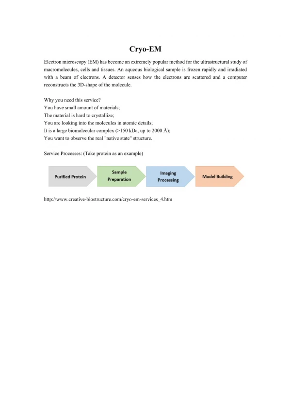
Electron microscopy (EM) has become an extremely popular method for the ultrastructural study of macromolecules, cells and tissues. An aqueous biological sample is frozen rapidly and irradiated with a beam of electrons. A detector senses how the electrons are scattered and a computer reconstructs the 3D-shape of the molecule.<br>

E N D
Cryo-EM Electron microscopy (EM) has become an extremely popular method for the ultrastructural study of macromolecules, cells and tissues. An aqueous biological sample is frozen rapidly and irradiated with a beam of electrons. A detector senses how the electrons are scattered and a computer reconstructs the 3D-shape of the molecule. Why you need this service? You have small amount of materials; The material is hard to crystallize; You are looking into the molecules in atomic details; It is a large biomolecular complex (>150 kDa, up to 2000 Å); You want to observe the real "native state" structure. Service Processes: (Take protein as an example) http://www.creative-biostructure.com/cryo-em-services_4.htm