Download
1 / 35
390 likes | 734 Vues
Neurotransmitter Actions. Direct action Neurotransmitter binds to channel-linked receptor and opens ion channels Promotes rapid responses Examples: ACh and amino acids. Neurotransmitter Actions. Indirect action

E N D
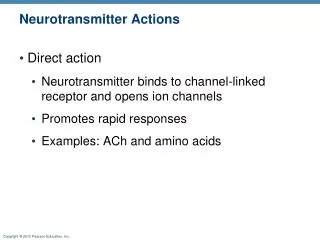
Neurotransmitter Actions • Direct action • Neurotransmitter binds to channel-linked receptor and opens ion channels • Promotes rapid responses • Examples: ACh and amino acids
Neurotransmitter Actions • Indirect action • Neurotransmitter binds to a G protein-linked receptor and acts through an intracellular second messenger • Promotes long-lasting effects • Examples: biogenic amines, neuropeptides, and dissolved gases
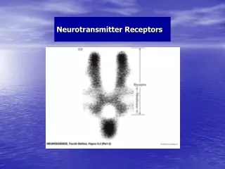
Neurotransmitter Receptors • Types • Channel-linked receptors • G protein-linked receptors
Channel-Linked (Ionotropic) Receptors • Ligand-gated ion channels • Action is immediate and brief • Excitatory receptors are channels for small cations • Na+ influx contributes most to depolarization • Inhibitory receptors allow Cl– influx or K+ efflux that causes hyperpolarization
Ion flow blocked Ions flow Ligand Closed ion channel Open ion channel (a) Channel-linked receptors open in response to binding of ligand (ACh in this case). Figure 11.20a
G Protein-Linked (Metabotropic) Receptors • Transmembrane protein complexes • Responses are indirect, slow, complex, and often prolonged and widespread • Examples: muscarinic ACh receptors and those that bind biogenic amines and neuropeptides
G Protein-Linked Receptors: Mechanism • Neurotransmitter binds to G protein–linked receptor • G protein is activated • Activated G protein controls production of second messengers, e.g., cyclic AMP, cyclic GMP, diacylglycerol or Ca2+
G Protein-Linked Receptors: Mechanism • Second messengers • Open or close ion channels • Activate kinase enzymes • Phosphorylate channel proteins • Activate genes and induce protein synthesis
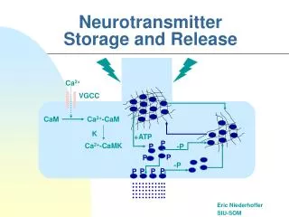
Closed ion channel Open ion channel 1 Neurotransmitter (1st messenger) binds and activates receptor. Adenylate cyclase Receptor G protein cAMP changes membrane permeability by opening or closing ion channels. 5a cAMP activates specific genes. 5c GDP 5b cAMP activates enzymes. 2 3 4 Receptor activates G protein. G protein activates adenylate cyclase. Adenylate cyclase converts ATP to cAMP (2nd messenger). Nucleus Active enzyme (b) G-protein linked receptors cause formation of an intracellular second messenger (cyclic AMP in this case) that brings about the cell’s response. Figure 11.17b
1 Neurotransmitter(1st messenger) bindsand activates receptor. Receptor (b) G-protein linked receptors cause formation of an intracellular second messenger (cyclic AMP in this case) that brings about the cell’s response. Figure 11.17b, step 1
1 Neurotransmitter(1st messenger) bindsand activates receptor. Receptor G protein GTP GDP GTP 2 Receptoractivates Gprotein. Nucleus (b) G-protein linked receptors cause formation of an intracellular second messenger (cyclic AMP in this case) that brings about the cell’s response. Figure 11.17b, step 2
1 Neurotransmitter(1st messenger) bindsand activates receptor. Adenylate cyclase Receptor G protein GTP GTP GDP GTP 2 3 Receptoractivates Gprotein. G proteinactivatesadenylatecyclase. Nucleus (b) G-protein linked receptors cause formation of an intracellular second messenger (cyclic AMP in this case) that brings about the cell’s response. Figure 11.17b, step 3
1 Neurotransmitter(1st messenger) bindsand activates receptor. Adenylate cyclase Receptor G protein ATP cAMP GTP GTP GDP GTP 2 3 4 Receptoractivates Gprotein. G proteinactivatesadenylatecyclase. Adenylatecyclase convertsATP to cAMP(2nd messenger). Nucleus (b) G-protein linked receptors cause formation of an intracellular second messenger (cyclic AMP in this case) that brings about the cell’s response. Figure 11.17b, step 4
1 Neurotransmitter(1st messenger) bindsand activates receptor. Adenylate cyclase Closed ion channel Open ion channel Receptor G protein 5a ATP cAMP changesmembrane permeability byopening and closing ionchannels. cAMP GTP GTP GDP GTP 2 3 4 Receptoractivates Gprotein. G proteinactivatesadenylatecyclase. Adenylatecyclase convertsATP to cAMP(2nd messenger). Nucleus (b) G-protein linked receptors cause formation of an intracellular second messenger (cyclic AMP in this case) that brings about the cell’s response. Figure 11.17b, step 5a
1 Neurotransmitter(1st messenger) bindsand activates receptor. Adenylate cyclase Closed ion channel Open ion channel Receptor G protein 5a ATP cAMP changesmembrane permeability byopening and closing ionchannels. cAMP GTP GTP 5b GDP GTP cAMP activatesenzymes. 2 3 4 Receptoractivates Gprotein. G proteinactivatesadenylatecyclase. Adenylatecyclase convertsATP to cAMP(2nd messenger). Nucleus Active enzyme (b) G-protein linked receptors cause formation of an intracellular second messenger (cyclic AMP in this case) that brings about the cell’s response. Figure 11.17b, step 5b
1 Neurotransmitter(1st messenger) bindsand activates receptor. Adenylate cyclase Closed ion channel Open ion channel Receptor G protein 5a ATP cAMP changesmembrane permeability byopening and closing ionchannels. cAMP GTP 5c GTP cAMPactivatesspecific genes. 5b GDP GTP cAMP activatesenzymes. 2 3 4 Receptoractivates Gprotein. G proteinactivatesadenylatecyclase. Adenylatecyclase convertsATP to cAMP(2nd messenger). Nucleus Active enzyme (b) G-protein linked receptors cause formation of an intracellular second messenger (cyclic AMP in this case) that brings about the cell’s response. Figure 11.17b, step 5c
Neural Integration: Neuronal Pools • Functional groups of neurons that: • Integrate incoming information • Forward the processed information to other destinations
Neural Integration: Neuronal Pools • Simple neuronal pool • Single presynaptic fiber branches and synapses with several neurons in the pool • Discharge zone—neurons most closely associated with the incoming fiber • Facilitated zone—neurons farther away from incoming fiber
Presynaptic (input) fiber Facilitated zone Discharge zone Facilitated zone Figure 11.21
Types of Circuits in Neuronal Pools • Diverging circuit • One incoming fiber stimulates an ever-increasing number of fibers, often amplifying circuits • May affect a single pathway or several • Common in both sensory and motor systems
Types of Circuits in Neuronal Pools • Converging circuit • Opposite of diverging circuits, resulting in either strong stimulation or inhibition • Also common in sensory and motor systems
Types of Circuits in Neuronal Pools • Reverberating (oscillating) circuit • Chain of neurons containing collateral synapses with previous neurons in the chain
Types of Circuits in Neuronal Pools • Parallel after-discharge circuit • Incoming fiber stimulates several neurons in parallel arrays to stimulate a common output cell
Patterns of Neural Processing • Serial processing • Input travels along one pathway to a specific destination • Works in an all-or-none manner to produce a specific response
Patterns of Neural Processing • Serial processing • Example: reflexes—rapid, automatic responses to stimuli that always cause the same response • Reflex arcs (pathways) have five essential components: receptor, sensory neuron, CNS integration center, motor neuron, and effector
Stimulus Interneuron 1 Receptor 2 Sensory neuron 3 Integration center 4 Motor neuron 5 Effector Spinal cord (CNS) Response Figure 11.23
Patterns of Neural Processing • Parallel processing • Input travels along several pathways • One stimulus promotes numerous responses • Important for higher-level mental functioning • Example: a smell may remind one of the odor and associated experiences
Developmental Aspects of Neurons • The nervous system originates from the neural tube and neural crest formed from ectoderm • The neural tube becomes the CNS • Neuroepithelial cells of the neural tube undergo differentiation to form cells needed for development • Cells (neuroblasts) become amitotic and migrate • Neuroblasts sprout axons to connect with targets and become neurons
Axonal Growth • Growth cone at tip of axon interacts with its environment via: • Cell surface adhesion proteins (laminin, integrin, and nerve cell adhesion molecules or N-CAMs) • Neurotropins that attract or repel the growth cone • Nerve growth factor (NGF), which keeps the neuroblast alive • Astrocytes provide physical support and cholesterol essential for construction of synapses
Cell Death • About 2/3 of neurons die before birth • Death results in cells that fail to make functional synaptic contacts • Many cells also die due to apoptosis (programmed cell death) during development