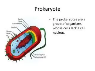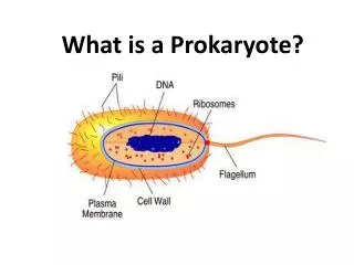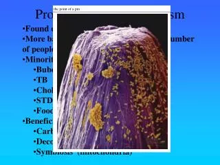
Prokaryote
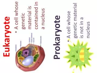
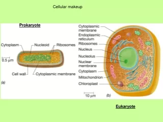
Cellular makeup. Prokaryote. Eukaryote. Table 1.2 Subgroups of Microorganisms. Perbedaan sel prokariot dan eukariot. Struktur organel sel Eukaryote (Starr, 1998). How do you study something that you cant see?. You look at it under the microscope

Prokaryote
E N D
Presentation Transcript
Cellular makeup Prokaryote Eukaryote
How do you study something that you cant see? • You look at it under the microscope • But certain microbes (e.g. bacteria) do not have too many identifying attributes • You grow large populations of them (i.e. culture them). • You observe the behavior of the population under various environmental conditions • On Solid media • In the presence of certain biochemicals • Based on the biochemical reactions they cause
magnification – ability to enlarge objects • resolving power – ability to show detail
Specimen preparation • wet mounts & hanging drop mounts – allow examination of characteristics of live cells: motility, shape, & arrangement • fixed mounts are made by drying & heating a film of specimen. This smear is stained using dyes to permit visualization of cells or cell parts.
Figure 3.8Preparing a hanging drop mount. Wet mounts (to observe living microbes) - Simple wet mounts : dry out quickly - hanging drop mounts : Using petroleum jelly to prevent drying of microbe samples
Stains - dyes that increase contrast by binding selectively to certain cells or to certain parts of them - vital stains : stain living cells, but more effective only after microbes have been fixed - Fixation : often distorts the cell’s appearance, motility can no longer be studied Heat fixation – using the open flame Chemical fixation – does less damage than heat ex) Osmic acid, formaldehyde, glutaraldehyde…..
Types of dyes - Basic dyes : with positive charges, stains the surfaces of most microbes - Acidic dyes : with negative charges, stains negatively charged parts of cells, including proteins ex) stain animal tissues that microbes have invaded - Mordants : intensify staining by increasing a cell’s affinity for a dye
Staining • cationic dyes - basic, with positive charges on the chromophore • anionic dyes - acidic, with negative charges on the chromophore • surfaces of microbes are negatively charged and attract basic dyes – positive staining. • negative staining – microbe repels dye & it stains the background
Staining • simple stains –one dye is used • differential stains – use a primary stain and a counterstain to distinguish cell types or parts. examples: Gram stain, acid-fast stain and endospore stain • special stains: capsule and flagellar stains
Preparing Smears for Staining • Stains consist of a positive and negative ion. • In a basic dye, the chromophore is a cation. • In an acidic dye, the chromophore is an anion. • Staining the background instead of the cell is called negative staining.
Staining procedures Simple stains – use basic dyes to make cells visible Differential stains – to distinguish between types of microoganisms 1. primary staining 2. Destaining 3. counterstaining ex) Gram stain (Gram positive and negative bacteria) Primary stain (Gentian violet) Mordant (Iodine) Decolorization (ethanol) Counterstain (safranin) Acid-fast stain (acid-fast bacteria : mycobacteria, some actinomycetes) Primary stain (Carbolfuchsin) Mordant (heating with steam) Decolorization (HCl + ethanol) Counterstain (Methylene blue)
Prosedur Pengecatan Gram • Pengecatan ini termasuk pengecatan positif-diferensial. Cuplikan inokulum harus diletakkan dalam gelas benda dan difiksasi dengan pemanasan. Beberapa reagen yang diperlukan adalah: • Crystal Violet (Primary Stain) • Iodine (Mordant) • Decolorizer (ethanol) • Safranin (Counterstain) • Aquades (dalam botol semprot)
Figure 3.9aGram-staining procedure.Steps of the procedure. • Special stains – reveal specific parts of a microbes, such as the wall, the nucleoid, an endospore, • membranes, flagella, or the capsule. • ex) flagella stains • renders flagella visible by adding a material, making them thicker (tannic acid) • thickened flagella are stained with dye (rosaniline) • negative stains • Identify the presence of capsules