Download
1 / 33
330 likes | 419 Vues
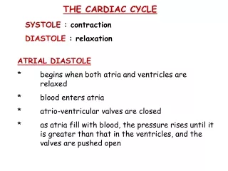
THE CARDIAC CYCLE. SYSTOLE : contraction DIASTOLE : relaxation. ATRIAL DIASTOLE * begins when both atria and ventricles are relaxed * blood enters atria * atrio-ventricular valves are closed

E N D
THE CARDIAC CYCLE SYSTOLE : contraction DIASTOLE : relaxation ATRIAL DIASTOLE * begins when both atria and ventricles are relaxed * blood enters atria * atrio-ventricular valves are closed * as atria fill with blood, the pressure rises until it is greater than that in the ventricles, and the valves are pushed open
ATRIAL SYSTOLE * when atrial diastole ends, the atria contract, pumping blood into the ventricles VENTRICULAR SYSTOLE * the ventricles then contract, causing a rise in pressure * the atrio-ventricular valves close - the first heart sound * the pressure forces open the semi-lunar valves, and the blood flows out
VENTRICULAR DIASTOLE * high pressure in the aorta and pulmonary artery forces blood back towards the ventricles, and this closes the semi-lunar valves : the second heart sound NB the pulse is caused by ventricular systole, and the elastic recoil of the arteries
THE STRUCTURE OF THE HEART • Why are the walls of the atria thinner than the walls of the ventricles? • Why is the wall of the right ventricle thinner than the wall of the left ventricle? • What happens to the atrioventricular valves when the ventricles contract, and why? • 4. What happens to the arterial valves when the ventricles relax, and why? • Which artery leaving the heart carries deoxygenated blood? • Which vein entering the heart carries oxygenated blood?
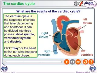
What is happening at 1, 2, 3 and 4? Annotate the diagram in your workbook
THE CONTROL OF THE HEART BEAT CARDIAC MUSCLE * branched * myogenic * does not fatigue
The Control of the Heartbeat Cardiac muscle contracts rhythmically on its own, but different areas have different intrinsic rates. The SINO-ATRIAL NODE (SAN) synchronizes the contraction of the different areas of cardiac muscle, so that the heart beats in a coordinated way.
The SAN sends out waves of excitation similar to nerve impulses across the surface of the atria, making them contract simultaneously. When these impulses reach the ATRIO-VENTRICULAR NODE (AVN), they travel down special cardiac muscle fibres called PURKYNE TISSUE, which fan out across the ventricles. These impulses cause the ventricles to contract in a wave, starting from the bottom.
An ECG This diagram shows the timing of the wave of excitation as it passes across the surface of the heart. An ELECTRO-CARDIOGRAM (ECG) shows this electrical activity.
TRANSVERSE SECTION THROUGH AN ARTERY Connective tissue Smooth muscle and elastic tissue Endothelium LUMEN
Remember its all about structure and function • Small lumen – for high pressure • Smooth endothelium – Reduce friction, and corrugated to allow expansion • Elastic fibres – allow for stretch and recoil • Smooth muscle – Allow for constriction of the artery if needed • Collagen fibres – To with stand the high pressure
* except the pulmonary artery, aorta, + except the pulmonary artery ^ except the pulmonary vein and carotid arteries
Arteries divide up into smaller ARTERIOLES, which divide to form capillary networks. Capillary networks drain into small veins called VENULES, which drain into large veins.
As well as a potential cause of blood clots, varicose veins can cause ulcerations of the legs. VARICOSE VEINS
DEEP VEIN THROMBOSIS (DVT) Blood clots that form in leg veins can dislodge and travel to other parts of the body, causing fatal problems such as heart attacks and pulmonary embolisms.
One cell think – allows for easy diffusion • Narrow lumen – allows only one blood cell down at a time, this slows the flow rate and allows time for diffusion
A CAPILLARY NETWORK – capillaries deliver food and oxygen to the tissues, at the same time removing carbon dioxide and other waste products. In order to do this efficiently, they are very narrow, with walls only one cell thick. This makes diffusion more efficient.
HOW DOES FLUID GET OUT OF THE CAPILLARY? Through pores in the capillary wall. WHICH COMPONENTS OF BLOOD WILL NOT PASS THROUGH THE PORES? Blood cells and large blood proteins.
CALIBRATING AN EYEPIECE GRATICULE Measure the size of the object using an eyepiece graticule. Record the size in graticule units. This is a stage micrometer: Each large unit on the micrometer is 1mm. Place this on the stage of the microscope with the graticule. Measure the actual length in mm of one graticule unit. Convert the size of the object from graticule units to mm.