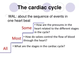
The cardiac cycle
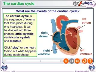
The cardiac cycle. In the normal heart, cardiac activity is repeated in a regular cyclical manner. With a resting heart rate of 75 beats/min, the duration of each cardiac cycle is 0.8 second. For the ventricles, the cycle consists of 0.3 second in systole and 0.5 second in diastole.


The cardiac cycle
E N D
Presentation Transcript
The cardiac cycle In the normal heart, cardiac activity is repeated in a regular cyclical manner. With a resting heart rate of 75 beats/min, the duration of each cardiac cycle is 0.8 second. For the ventricles, the cycle consists of 0.3 second in systole and 0.5 second in diastole.
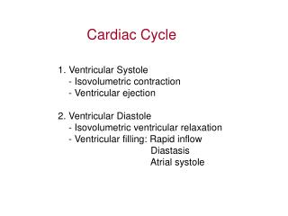
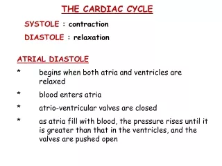
The cardiac cycle * Atrial systole, * Ventricular systole, then * Diastole of the whole heart Atrial and ventricular systoles do not occur at the same time, but their diastoles occur at the same time during the diastole of the whole heart.
The cardiac cycle * Atrial systole * Ventricular systole Isometric (isovolumetric) contraction phase Rapid ejection phase Slow ejection phase * Diastole of the whole heart Protodiastole Isometric (isovolumetric) relaxation phase Rapid filling phase Slow filling phase
LATE DIASTOLE DIASTOLE ATRIAL SYSTOLE VENTRICULAR EJECTION ISOMETRIC VENTRICULAR CONTRACTION
EJECTION ISOVOLUMETRIC CONTRACTION ISOVOLUMETRIC RELAXATION RAPID INFLOW DIASTASIS ATRIAL SYSTOLE AORTIC PRESSURE PRESSURE (mmHg) ATRIAL PRESSURE VENTRICLE PRESSURE VOLUME (ml) ECG PHONO- CARDIOGAM SYSTOLE DIASTOLE SYSTOLE
CARDIAC CYCLE Reduced Ejection Reduced Ventricular Filling Rapid Ventricular Filling Rapid Ejection Isovolumic Relax. Atrial Systole Atrial Systole Isovolumic contract. Aortic opens Aortic closes Mitral opens Mitral Closes S1 S2
Heart Sounds • Outline: • Audible and inaudible heart sounds. • First and second heart sounds: • - Relation to cardiac cycle • - Causes, characters, site of hearing & significance • Third and Fourth heart sounds: • - Relation to cardiac cycle • - Causes, characters, site of hearing & significance • Further Reading: • Guytin: Textbook of Medical Physiology • Ganong: Review of Medical Physiology
HEART SOUNDS • During each cardiac cycle 4 heart sounds are produced due to physiological vibration in the heart: • The 1st & 2nd heart sounds are audible i.e. normally heard when a stethoscope is applied to the chest wall. • The 3rd heart sound is usually inaudible (different to be heard); however, it can be sometimes heard in some children. • The 4th heart sound is normally inaudible.
The heart sounds The first heart sound occurs at the beginning of systole and sounds like the word LUP. The second heart sound occurs at the beginning of diastole and sounds like the word DUP. The interval between the first and second sound is shorter than the interval between the second and the next first heart sound.
FIRST HEART SOUND • It occurs at the beginning of ventricular systole i.e. isometric contraction phase and it extends into the phase of maximum( rapid ) ejection. • Causes: • 1) Sudden closure of the AV valves ( mitral & tricuspid ) at the start of the ventricular systole which produces vibrations in the cusps of the AV valves (valvular factor).
2) Contraction of the ventricular muscle which causes vibrations in the muscle itself and vibration in the chorda tendinae ( ventricular) factors. 3) Ejction of blood from the venticles into the aorta and pulmonary artery which causes vibrations in these walls ( vascular factors). • Charcters • Soft and law pitched • It’s duration is about 0.15 second. • It is heard as the ward ((lup)), by the stethoscope.
Site of hearing: • - it is best heard at the mitral area= 5th left intercostl space about 10 cm from the midline • Tricuspid area= lower part of the sternum (at the 6th right stenocostal junction). • Significance: 1)it indicates the start of ventricular systole. 2)It gives an idea about the condition of the AV valves. 3) Its intensity and duration indicates the condition of the cardiac muscle (Myocardium).
2) Second heart sound: • It occurs at the onset of ventricular diastole isometric relaxation phase of the cardiac circle -Causes: Sudden closure of the semi lunar valves ( aortic and pulmonary valves) and the vibrations that follow in their cusps (= valvular) factors. vibration in the walls of the aorta and pulmonary artery due to vibration in the aortic and pulmonary valves (= Vascular factors).
-Characters • Sharp and high pitched • Its duration is about 0.12 seconds • Its heard as the spoken ward “Dup” by the stethoscope • Site of hearing: • It is best heard at: Aortic area ( = 2nd right stennocostal junction). Pulmonary area ( = 2nd left intercostal space near the sternum).
-Significance: It indicates the end of ventricular systole and the start of ventricular diastole It gives an idea about the condition of the semi lunar valves. Its intensity is directly proportional with the arterial blood pressure. -N.B. The duration between the 1st and 2nd heart sounds represents clinically the time of ventricular systole and that between the 2nd & 1st heart sounds as the time of ventricular diastole.
THIRD HEART SOUND • It occurs during ventricular diastole; during maximum • (rapid) filling phase of ventricular diastole. So, it comes • after the 2nd heart sound. • Causes: • vibrations of the relaxed ventricular walls due to rush of blood from the atria into the ventricles during rapid ventricular filling. • Characters: • Soft and low pitched • Its duration is about 0.1 second. • It is usually inaudible. It can be sometimes heard in children or persons with very thin chest wall. • Significance: • it indicates opening of the AV valves during ventricular diastole and rapid ventricular filling.
FOURTH HEART SOUND • It occurs during the phase of atrial systole; therefore, • it is called atrial heart sound. • Causes: • Vibrations in the relaxed ventricular walls due to rush of blood from the atria into the ventricles under the effect of atrial contraction. • Characters: • It is a faint low pitched sound. • Its duration is about 0.5 second. • It is normally inaudible but can be recorded or detected by the phonocardiogram. • Significance: • it indicates atrial systole (=late ventricular filling = late ventricular diastole).
MURMURS • Cardiac murmurs are abnormal heart sounds in the form of noises resulting from turbulent blood flow, • Murmurs may be: • Systole murmurs, if they occur during systole. • Diastolic murmurs, if they occur during diastole • Causes: • The main factors causing cardiac murmurs include: • Stenosis (narrowing) of heart valves • Incompetence of heart valves • Increase of blood flow or decrease of blood viscosity (as in anemia).
Pressure changes during the cardiac cycle • Outline: • Aortic pressure changes during the cardiac cycle • Arterial pulse curve (radial pulse curve). • The arterial pulse and its importance. • The pulse pressure and water-hammer pulse. • Atrial pressure changes during the cardiac cycle. • The jugular venous pulse waves • Jugular pulse curve and its importance. • Further Reading: • Guyton: Textbook of Medical Physiology • Ganong: Review of Medical Physiology
PRESSURE CHANGES DURING THE CARDIAC CYCLE • AORTIC PRESSURE CHANGES • At the beginning of the ventricular systole and when the aortic valves opens (i.e. during the maximum or rapid ejection phase of the cardiac cycle), the aortic pressure is rapidly increases to reached a maximum called systolic blood pressure (about 120 mm Hg). • During the reduced ejection phase and the protodiastole phase, there is a fall in the aortic pressure due to slow ejection and the flow of blood from the aorta into the peripheral arteries. • During ventricular diastole, there is a gradual decrease of aortic pressure due to continued flow of blood from the aorta into the systemic peripheral vessels. The aortic pressure decreases to a diastolic valve of about 80 mmHg.
The arterial pressure wave = (Radial pulse curve) • This reflects the aortic pressure changes during the cardiac cycle. • Recording of the arterial pressure wave (using a special apparatus called sphygmograph), gives a curve which consists of an ascending limb called the anacrotic limb and a descending limb called the catacrotic limb. • The anacrotic limb is due to sudden distension (expansion) of the artery during ejection of blood from the left ventricle (i.e. during ventricular systole). • The catacrotic limb is due to flow of blood from the artery into more peripheral vessels during the late part of ventricular systole and during ventricular diastole. • Over the catacrotic limb, there is an irregularity in the form notch (called diacrotic notch or incisura) followed by a wave (called dicrotic wave).
- The diacrotic notch occurs during closure of the aortic valves at the end of the protodiastolic phase (=beginning of the ventricular diastole). • The diacrotic wave occurs during ventricular diastole (isometric relaxation phase) and it is due to the elastic recoil of the arterial wall. • THE ARTERIAL PULSE • During ventricular systole (rapid ejection phase), the expansion of the aortic wall which is rapidly propagated through the arterial walls can be felt using the middle three fingers as a pulsation. • Palpitation of the radial pulse (=arterial pulse) gives information about: • Heart Rate; its regularity, the presence of missed beats or extra systoles. • a
The force of ventricular contraction e.g. full pulse in muscular exercise and weak pulse in hemorrhage. • The condition of the arterial wall which indicates the arterial elasticity e.g. normal arterial wall is soft and in arteriosclerosis it is rigid. • PULSE PRESSURE • Pulse pressure is the difference between systolic blood pressure and diastolic blood pressure. • It increases in conditions that increase systolic BP and/ or decrease the diastolic BP e.g. • Aortic regurgitation. • Arteriosclerosis • big pulse pressure produces marked arterial pulsations (water-hammer pulse).
N.B: • Pressure changes in the pulmonary artery during the • cardiac cycle are similar to those in the aorta but they differ • in magnitude (the systolic BP is 25-30 mm Hg and the • diastolic BP is 8-10 mm Hg). • ATRIAL PRESSURE CHANGES • During each cardiac cycle, atrial pressure is increased during the following phases: • Atrial systole • Isometric (isovolumetric) contraction of the ventricles due to bulging of the AV valves towards the atria which results from increased ventricular pressure. • Protodiastolic phase and isometric relaxation of the ventricles due to accumulation of blood in the atria while the AV valves are closed.
JUGULAR VENOUS PULSE WAVES = (Jugular pulse curve) • The pressure changes in the right atrium during the cardiac cycle are transmitted to the external jugular vein producing similar changes in the venous pressure of this vein venous pulse waves. • This is because there are no valves between the vena cava and the right atrium. • The jugular venous pulse waves can be recorded using a special apparatus called phlebograph. • The record shows 3 positive waves (a, c, v) and 3 negative waves (x, x1, y): • The a-wave is due to atrial systole which causes increase in the intra-atrial pressure. • The x-wave is due to atrial diastole.
The C-wave occurs at the beginning of the ventricular systole i.e. during the isometric contraction of the ventricles. • It is due to bulging of the tricuspid valve towards the right atrium which causes increase of the right atrial pressure (which is conducted to the external jugular vein). • The X-wave is due to downward displacement of the A-V valve (tricuspid valve) and the AV ring during maximum ejection phase of ventricular contraction. • The V-wave is due to accumulation of venous blood in the right atrium increase of atrial pressure during the late part of the ventricular systole (protodiastolic phase) and also during the isometric relaxation of the ventricles.
The Y-wave is due to opening of the AV valve (tricuspid valve) during maximal ventricular filling phase i.e. rush of blood from the atria into the ventricles decrease of atrial pressure. • SIGNIFICANCE OF THE JUGULAR • VENOUS PULSE CURVE • The “a-c” interval represents conductivity in the S-V bundle. • - The “a-c” interval is measured from the beginning of the a-wave to the beginning of the c-wave. • - Since the a-wave is due to atrial systole and c-wave is due to ventricular systole, the a-c interval measures • the time of conduction of the cardiac impulse from the • right atrium to the ventricles. • - The a-c interval corresponds to the P-R interval of the ECG. It is normally about 0.12 second.
- The “a-c” interval is prolonged in cases of delayed conductivity in the AV bundle which is an early sign of heart block. • In partial heart block, the number of “a” waves is greater than the number of the “c” or “v” waves. • In atrial fibrillation, the “a” wave is absent. • N.B: • When the jugular pulse curve is simultaneously recorded with the radial pulse curve, this is called polygram. • The wave preceding the ascending limb of the radial pulse curve is the c-wave.
CARDIAC CYCLE Reduced Ejection Reduced Ventricular Filling Rapid Ventricular Filling Rapid Ejection Isovolumic Relax. Atrial Systole Atrial Systole Isovolumic contract. Aortic opens Aortic closes Mitral opens Mitral Closes S1 S2