Download
1 / 22
220 likes | 329 Vues
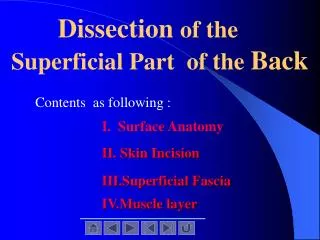
Dissection of the Superficial Part of the Back. Contents as following :. I. Surface Anatomy. II. Skin Incision. III.Superficial Fascia. IV.Muscle layer. Place the cadaver in the prone position.

E N D
Dissection of the Superficial Part of the Back Contents as following : I. SurfaceAnatomy II. Skin Incision III.Superficial Fascia IV.Muscle layer
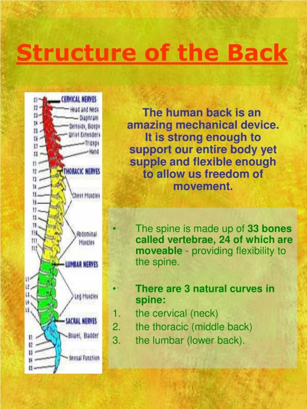
I. SurfaceAnatomy:Palpate the following bony landmarks: 1 the external occipital protuberance, 2 the vertebra prominens (spinous process of C7), 3 spinous process of first thoracic vertebra, 4 the crest of the spine of the scapula, 5 the acromion process of the scapula, 6 the superior and inferior angles of the scapula, 7 the iliac crest.
II. Skin Incision: 1. Make a vertical midline skin incision from the external occipital protuberance downward to the level of the iliac crest. 2. At the lower end,extend the incision transversely to the right and left as far as themidaxillary line. 3. At the upper end, extend the incision from the external occipital protuberance to the acromion process. 4. On the right and left sides, reflect the skin laterally from the back of the neck, the shoulders, and the back to the midaxillary lines.
III. Superficial Fascia (subcutaneous tissue) :Identify the cutaneous branches of the posterior rami of the cervical,thoracic,and lumbar spinal nerves in the underlying subcutaneous tissue.
1. Locate the greater occipital nerve, which is the posterior ramus of the second cervical nerve. It emerges from thetrapezius about 2.5cm below andlateral to the external occipital protuberance.2. Clean and study one cutaneous branch of a posterior ramus of a thoracic spinal nerve.3. To assist you in findingsuch a nerve, carefully look for a blood vessel that follows each of these nerves. Do not waste time tracing out the cutaneous branches of all the posterior rami
Now reflect the superficial fascia of the back in line with the skin incisions, being carefully not to damage the underlying muscles, the trapezius and the latissimus dorsi.
IV. Muscle: Identify and clean the trapezius and latissimus dorsi muscles and origin of the posterior part of the deltoid muscle
Trapezius muscle. Confirm that this muscle has an extensive origin from the medial one-third of the superior nuchal line of the occipitl bone, the occipital protuberance, the ligamentum nuchae, the spinous processes of the seventh cervical vertebra and the spinous processes and supraspinous ligaments of thoracic vertebrae one to twelve.
Trace the superior muscle fibers to theirinsertion on the lateral third of the clavicle, the middle fibers to their insertion on the acromion and the spine of the scapula, and the inferior fibers to their insertion on the base of the spine of the scapula.
Cut free the origin of the trapezius from the skull, the ligamentumnuchae, and the spinous processes of the vertebrae. Carefully reflect the trapezius laterally, taking care not to damage the underlying levator scapulae and rhomboid muscles.
Identify and preserve the spinal part of the accessory nerve and the posterior rami of the third and fourth cervical nerves, which supply the trapezius muscle.
Latissimus dorsi muscle.Confirm that this muscle arises from the posterior part of the iliac crest, the lumbar fascia, the spinous processes of the lower six thoracic vertebrae, and from the lower three or four ribs; sometimes a few fibers arise from the inferior angle of the scapula. Note that the fibers sweep upward and laterally, converging on a tendon that passes into the axilla to be inserted into the humerus.
Transect the origin of the latissimus dorsi and reflect the muscle laterally, thus exposing the underlying serratus posterior inferior muscle.
Identify and clean the levator scapulae and rhomboid minor and rhomboid major muscles. Leave the levator scapulae intact and trace its fibers to their insertion on the medial border of the scapula.
Cut free the origins of the rhomboid minor and major muscles from the vertebrae and carefullyreflect them laterally to their insertions on the medial border of the scapula. Be careful to preserve the underlying serratus posterior superior muscle, which is sometimes stuck to the rhomboid muscles.
Identify and clean the dorsal scapular nerve and the deep branches of the superficial cervical vessels that lie deep to the rhomboid muscles, close to their insertion.
Identify the serratus anterior muscle at its insertion along the anterior surface of the medial border of the scapula.
Define the posterior border of the deltoid muscle and confirm the origin of this muscle from the spine and acromion process of the scapula. With a scalpel, cut free the origin of the deltoid muscle from the spin and acromion process but leave its attachment to the clavicle intact. Note that the deltoid fibers pass downward to their insertion on the deltoid tuberosity of the humerus. To assist you with the later study, now remove the skin and superficial fascia from the back of the shoulder and the back of the arm, down as far as the olecranon process of the ulna.
Identify and clean the supraspinatus, infraspinatus,teres minor and teres major, and the long head of the triceps muscles.Expose the boundaries and contents of the quadrilateral and triangular spaces.