Chest Injuries: Mechanisms, Treatment & Assessment
460 likes | 705 Vues
Understand chest injuries, ventilation mechanics, signs & symptoms, and life-saving treatments. Learn how to assess and provide initial interventions for chest trauma in emergency situations.

Chest Injuries: Mechanisms, Treatment & Assessment
E N D
Presentation Transcript
Mechanics of Ventilation(1 of 2) • Inspiration • Intercostal muscles contract and diaphragm flattens. • Expiration • Intercostal muscles and diaphragm relax; tissues move back to normal position.
Mechanics of Ventilation(2 of 2) • Phrenic nerves exit the spinal cord at C3, C4, and C5. • Spinal cord injury below C5 • Loss of ability to move intercostal muscles • Diaphragm can still contract; patient can still breathe. • Spinal cord injury at C3 or higher • No ability to breathe
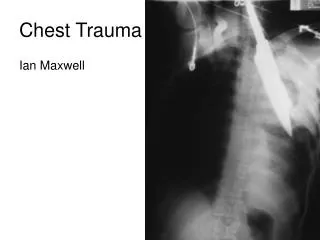
Injuries to the Chest • Closed chest injuries • Caused by blunt trauma • Open chest injuries • Caused by penetrating trauma
Pain at site of injury Pain aggravated by increased breathing Bruising to chest wall Crepitus with palpation of chest Penetrating injury to chest Dyspnea Hemoptysis Failure of chest to expand normally Rapid, weak pulse and low blood pressure Cyanosis around lips or fingernails Signs and Symptoms
You are the Provider • You and your EMT-B partner are dispatched to a construction site where a worker fell on a piece of metal and has an open chest wound. • You arrive and see no scene hazard or need for other resources.
You are the provider continued • What is the mechanism of injury? • What precautions should you take?
Scene Size-up • Observe for hazards. • Do not disturb potential evidence. • Put several pairs of gloves in your pocket. • Consider spinal immobilization. • Ensure that police are on scene if incident involved violence.
You are the provider continued (1 of 2) • Your partner provides spinal immobilization. • The patient is responsive with an open airway. He says, “I can’t catch my breath. It hurts.” • You find a penetrating injury on the right anterior portion of the chest. • You see a small amount of bleeding. The blood bubbles as your patient breathes.
You are the provider continued (2 of 2) • What are the steps of the initial assessment? • What life-saving treatments would you provide? • What treatment do you provide? • Describe the rest of your assessment process after the initial assessment.
Initial Assessment • General impression • Quickly evaluate ABCs. • Difficulty speaking may indicate several problems. • Patients with significant chest injuries will look sick. • Airway and breathing • Ensure that patient has a clear, patent airway. • Protect the spine. • Inspect for DCAP-BTLS.
Inspection • Decreased breath sounds usually indicate significant damage to a lung. • If both sides of chest do not have equal rise and fall, chest muscles have lost ability to work properly. • If one section of chest moves in opposite direction from the rest of the chest (paradoxical motion), this is a life threat.
Immediate Interventions • Apply an occlusive dressing to any penetrating chest injury. • Stabilize paradoxical motion with a large bulky dressing and 2'' tape. • Apply oxygen via nonrebreathing mask at 15 L/min. • Provide positive pressure ventilations if breathing is inadequate.
Circulation • Assess patient’s pulse. • Consider aggressive treatment for shock. • Internal bleeding can quickly cause death.
Transport Decision • Rapidly transport if patient has problems with ABCs. • Pay attention to subtle clues. • Skin signs • Level of consciousness • Sense of impending doom
You are the provider continued • You ensure that his airway is open. • Breathing is labored; you start oxygen via nonrebreathing mask at 15 L/min. • You quickly assess for DCAP-BTLS; seal sucking chest wound with an occlusive dressing. • You check distal pulse. Skin is clammy. Bleeding is noted and controlled. • High-priority transport
Focused History and Physical Exam • Focused physical exam • For a patient with isolated chest injury and limited MOI • Rapid physical exam • For a patient with a significant MOI • Use DCAP-BTLS. • Do not focus just on the chest wound. • Obtain baseline vital signs. • Obtain SAMPLE history quickly.
Interventions • Provide complete spinal immobilization. • Maintain open airway; be prepared to suction. • Provide assisted ventilations if needed. • Control bleeding. • Place occlusive dressing over penetrating chest wound. • Stabilize flail segment with a bulky dressing. • Treat aggressively for shock. • Do not delay transport.
Detailed Physical Exam • Perform en route to the hospital if time allows.
Ongoing Assessment • Assess effectiveness of interventions. • Reassess vital signs. • Communication and documentation • Communicate with hospital early if patient has significant MOI. • Describe injuries and treatment given.
You are the provider continued • Patient has a significant MOI; do a rapid physical exam. • Obtain baseline vital signs and SAMPLE history. • Take c-spine precautions and transport continuing oxygen therapy. • Perform detailed physical exam and ongoing assessment en route.
Complications of Chest Injuries A pneumothorax occurs when air leaks into the space between the pleural surfaces.
Pneumothorax • Air accumulates in the pleural space. • Air enters through a hole in the chest wall. • The lung may collapse in a few seconds or a few minutes. • An open or penetrating wound to the chest is called a sucking chest wound.
Care for Open Pneumothorax • Clear and manage the airway. • Provide oxygen. • Seal an open wound with an occlusive dressing. • Depending on local protocol, tape down all four sides or create a flutter valve.
Spontaneous Pneumothorax • Some people are born with or develop weak areas on the surface of the lungs. • Occasionally, the area will rupture spontaneously, allowing air into the pleural space. • Patient experiences sudden chest pain and trouble breathing. • Consider a spontaneous pneumothorax for a patient with chest pain without cause.
Tension Pneumothorax (1 of 2) • Can occur from sealing all four sides of the dressing on a sucking chest wound • Can also occur from a fractured rib puncturing the lung or bronchus • Can also result from a spontaneous pneumothorax
Signs and Symptoms ofTension Pneumothorax • Respiratory distress • Distended neck veins • Tracheal deviation • Tachycardia • Low blood pressure • Cyanosis • Decreased lung sounds
Care for Tension Pneumothorax • If a tension pneumothorax develops from sealing an open chest wound, partly remove the dressing to let the air escape. • If there is no open wound, follow local protocol.
Hemothorax (1 of 2) • Collection of blood in the pleural space • Suspect if the following are seen: • Signs and symptoms of shock • Decreased breath sounds on affected side • If both air and blood are present in the pleural space, it is a hemopneumothorax.
Rib Fractures • They are very common in the older people. • A fractured rib may lacerate the surface of the lung. • Patients will avoid taking deep breaths and breathing will be rapid and shallow. • The patient often holds the affected side to minimize discomfort. • Administer oxygen.
Flail Chest (1 of 2) • Segment of chest wall detached from rest of thoracic cage • Occurs when: • Three or more ribs are fractured in two or more places. • Sternum is fractured along with several ribs. • Creates paradoxical motion
Care for Flail Chest • Maintain airway. • Provide respiratory support with BVM if needed. • Perform ongoing assessments for pneumothorax and other respiratory complications. • Immobilize flail segment.
Pulmonary Contusions • Bruising of the lung • Develops over hours • Alveoli fill with blood, and edema accumulates in the lung, causing hypoxia. • Provide oxygen and ventilatory support.
Traumatic Asphyxia • Sudden, severe compression of chest • Produces rapid increase in pressure within chest • Results in neck vein distention, cyanosis, and bleeding into the eyes • Provide supplemental oxygen and monitor vital signs. • Transport immediately.
Blunt Myocardial Injury • Bruising of heart muscle • Pulse is often irregular. • There is no prehospital treatment for this condition. • Check patient’s pulse and note irregularities. • Provide supplemental oxygen and transport immediately.
Pericardial Tamponade (1 of 2) • Blood or other fluids collect in the pericardium.
Pericardial Tamponade (2 of 2) • Signs and symptoms: • Very soft and faint heart tones • Weak pulse • Low blood pressure • Decrease in difference between systolic and diastolic blood pressure • Jugular vein distention (JVD) • Provide oxygen and transport quickly.
Laceration of the Great Vessels • The superior vena cava, inferior vena cava, pulmonary arteries and veins, and aorta are contained in the chest. • Injury to these vessels can cause fatal hemorrhage. • Treatment includes: • CPR • Ventilatory support • Supplemental oxygen • Transport immediately.