Download
1 / 1
10 likes | 149 Vues
ISOLATION AND CHARACTERIZATION OF NOVEL MICROBACTERIOPHAGE BARBARA Angel Mitchell, Department of Biological Sciences. College of Arts and Sciences, and Honors College Faculty Mentors: Stephanie Simon, Robert Benjamin, and Lee Hughes,

E N D
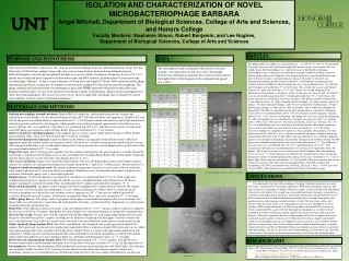
ISOLATION AND CHARACTERIZATION OF NOVEL MICROBACTERIOPHAGE BARBARA Angel Mitchell, Department of Biological Sciences. College of Arts and Sciences, and Honors College Faculty Mentors: Stephanie Simon, Robert Benjamin, and Lee Hughes, Department of Biological Sciences, College of Arts and Sciences RESULTS PURPOSE AND HYPOTHESIS DATA The Honors Hall soil sample was collected from 33° 12’266”N, 97° 09’115”W underneath a tree. Bird feces was not collected in sample but found in vicinity of extraction. Soil was slightly damp. The bacteriophage was enriched to increase likelihood of obtaining bacteriophage from environment versus prospect of plaques with direct plating. After first plating, positive plate was completely clear, negative plate was covered with bacteria, and plate with four test spots each showed positive plaques. The spot test objective was determination of phage growth. After bacterial lawn, serial dilutions were performed on sample from first enrichment. The first phage titer assay was performed on the 10-3 plate that had 27 plaques and incubated at 37°C for 24 hours. The second titer assay was performed from the 10-2 plate and incubated at 37°C for 24 hours. The second attempted assay purification had no plaques on the plates. As the second assay was unsuccessful, the 2nd titer assay was performed again from the 10 -2 plated and incubated at 37°C for 36 hours. The results from the successful third assay were as follows: The positive control contained four 0.5 cm diameter phage, 10-1 plate contained about 500 plaques, 10-2 plate contained about 100 plaques, 10-3 plate contained 5 plaques, and 10-4 plate contained no visible plaques. 100 plate had too many plaques to count and was used for flooding. The 100 plate was flooding with phage buffer. The PB was aspirated from the plate and placed into conical tube. The phage lysate spot test was performed from 100 to 10-10 with 10 minutes of M. smegmatis infection and left at 37°C for 24 hours of incubation. The phage titer assay was calculated utilizing the 10-7 plate. The plaque area was calculated as π and plate area was (42.5 mm)2 • π. pfu(max web) = 1806.25 π/π =1806.25 mm2. Titer lysate calculation: 1 pfu/1 μL • 100 μL/1 mL • 107 = 2.0 • 109 pfu/ml Phage stock was calculated to determine dilutions and volumes required to form a desired web pattern on bacterial lawn grown on top agar.V = 2.0 • 106 pfu/μL. Necessary dilution was calculated to be optimal at 10 μL per plate. Serial dilution was then performed with 10-4 from 10-1 to 10-4, and infected with M. smegmatis. Samples then plated and incubated. The goal of the 10-plate phage infection and harvest was to obtain a high-titer phage lysate with high enough phage concentration for progression to DNA-isolation. The PB plates were flooded then harvested into the conical tube and stored at 4°C. Serial dilutions on lysate harvest were performed from 100 to 10-11 and plated from 10-6 to 10-11, then incubated. Results from the serial dilution plates were shown on 10-6 plate with about 140 plaques. The 10 mL lysate harvest was transferred to 50 mL Oakridge tube and remaining lysate saved at 4°C. 40 μL Nuclease was added to tube, incubated and cooled. 4 mL phage precipitate solution was added and mixed by gentle inversion, then incubated at 37°C for 24 hours. High Titer Lysate was calculated as 1.4 • 1010 pfu/mL. Grids were prepared and sent for electron microscopy. After DNA isolation and purification, 49 μL DNA solution had accumulated. The DNA concentration was 240.80 ng. 241 ng/μL • 49 μL = 11809 ng of total recovered DNA. Electrophoresis was performed for various restriction enzymes. This project involved the isolation of a new strain of mycobacteriophage from soil collected in Denton, Texas near the University of North Texas campus using a Mycobacterium smegmatis host strain and an enrichment protocol. Individual plaques were selected and purified through 4 successive rounds of isolation. A high titer lysate of 2.0 x 10 9 pfu/ml was created and used to perform electron microscopy and DNA isolation. Transmission electron microscopy revealed phage “Barbara” to have a capsid diameter of 50 nm and a tail length of 150 nm. DNA restriction of the phage chromosome produced a unique gel electrophoresis pattern when compared to other known mycobacteriophage. This phage isolation and characterization was performed as part of the HHMI-sponsored National Genomics Research Initiative and the isolate was one of 24 obtained by freshmen students in the program. Many mycobacateriophage also infect the related pathogenic Mycobacterium tuberculosis, and it is hoped that such phage may eventually be used to treat antibiotic resistant strains of this human pathogen. The electrophoresis gels, performed with various restriction enzymes, were measured at each cut to show relationship between size and distance migrated. For each restriction enzyme, the length of the visible fragments were estimated and placed into a table. MATERIALS AND METHODS Isolating bacteriophage through enrichment. Honors Hall soil sample was collected and collection site location and characteristics noted. Sample was transferred to Erlenmeyer flask; H20, 7H9 Glycerol Broth, AD supplement, 100mM CaCl2 and host M. Smegmatis were added. Flask was then incubated at 37°C for 24 hours contents transferred to conical tube and bacterial cell and large debris collected by centrifugation. Phage particles were collected from the supernatant by ultra filtration into a micro-centrifuge tube. A serial dilution of the filtrate was performed from 100 to 10-4 and plated, with a positive control made using D29 phage and a negative control of Phage Buffer. Plates are incubated at 37°C for 24 hours. Spot test on putative enrichment plaques. A micropipette tip was used to transfer sample putative plaques to PB in labeled microcentrifuge tubes, which were then vortexed and set aside for re-plating. Bacterial lawn prepared in top agar. A grid was drawn on the bottom of an agar plate with individual sectors labeled for appropriate phage destinations and positive/negative controls. The plate was covered with top agar inoculated with host bacteria. After top agar solidification, 5 μL of each putative phage isolate was spotted onto the correspondingly labeled grid location. Plate was inverted and incubated at 37°C overnight. Phage-Titer assay. After verifying putative plaques from the original isolation plates, the spot test phage was further purified by transferring a small plug from the plaque area to a microcentrifuge tube containing Phage Buffer. The resultant phage suspension from the chosen plaque was then titered by serial dilution from 10-1 to 10-10. Titer Assay Calculation. Plaques were counted for dilution plates with 20 to 200 plaques/plate and recorded. Plaque forming units per mL (pfu/mL) are calculated according to the formula: titer(pfu/mL) = (pfu/# μL) • (1000 μL/mL) • dilution factor (10-x) Empirical calculation and phage stock. The number of plaque forming units necessary to create a max web was calculated using formula: pfu(max web) = area (agar plate)/ area (plaque). Dilution necessary for infection with optimal web pattern was calculated. Volume(mL) phage stock = (desired pfu)/(pfu/mL) Empirical assay. 10 fold serial dilution as determined by calculations was performed from 10-1 to 10x. Each sample tube, including negative/positive controls were infected with M. smegmatis and diluted. Phage infected bacteria for 25 minutes. Samples and negative control were plated. Plates sat undisturbed for 35 minutes and incubated overnight at 37°C. Phage stock preparation. The phage stock was prepared by harvesting phage from 10 plates infected with PB. The volume necessary for a web on one plate was multiplied by 10 to get volume needed for all 10 plates. Flask was infected with M. smegmatis and phage was infected for same amount of time as empirical test. 55°C top agar was added to infected M. smegmatis then plated onto all 10 plates. Control plate was prepared. Upon TA set, plate was incubated at 37°C overnight. 10 Plate phage harvest. After phage stock was prepared, the ten plates were flooded with phage buffer for several hours. The phage buffer was collected into a conical tube and centrifuged for 20 minutes to pellet cell debris. Supernatant was collected and transferred into filter-sterilization unit. Phage Titer.Serial dilutions performed on lysate. 10 μL each dilution from 10-6 to 10-11, positive, negative control was added to M. smegmatis and left for 25 minutes, then plated. Next day, plaques were counted to determine if enough DNA for purification. Electron Microscopy. Parafilm, Petri dish lid, and grids utilized. Micropipettor was used to place phage preparation onto grid. Phage was absorbed onto grid for 2 minutes according to titer. Fluid was wicked up with filter paper. Grid was washed twice, once with sterile water, once with 1.0% uranyl acetate, then with stain, then allowed to air-dry and was stored for microscopy. Isolate and purify phage genomic DNA. After final serial dilutions were incubated, tube is high-speed centrifuged for 20 minutes. The supernatant was decanted, leaving the pellet undisturbed. Excess liquid was drained. DNA clean-up resin was added to precipitate phage pellet and resuspended. Resin placed into column. Waste was removed through column and flushed with isopropanol. Column transferred to microcentrifuge tube and centrifuged for one minute, then phage was eluted from column, elution buffer then added to resin and centrifuged. DNA was combined, centrifuged, then stored at 4°C overnight. Restriction and analyzing phage genomic DNA. DNA mixed and incubated at 65°C for 10 minutes, then placed on ice. Reactions represented in predetermined calculations were set up. Tubes were spun, incubated 37°C for 2 hr, then placed on ice. Electrophoresis. Gel was already prepared. DNA standard was created in microcentrifuge tube with DNA ladder, 10x restriction enzyme buffer, and H2O to make 10 μL. Tracking dye was added to each reaction tube and mixed gently. Samles were immediately placed on ice. A micropipettor tip was utilized to load gel with 10 μL each sample. Electrophoresis was then ran. UncutDNA, BamHI, ClaI, Ladder, EroRI, HaeI, HindI CONCLUSIONS Considering the two types of bacteriophage, the phage from this experiment was most likely lytic, considering the clear plaque appearance. With either type plaque, there are four steps to infection. Adsorption of phage to bacteria’s surface is done with tip of tail that adheres to bacterial cell receptors. The second step, irreversible attachment of phage is practically an extension of adsorption minus specificity. Penetration of bacterial wall, the third step, occurs due to contraction of bacterial sheath forming something like a vacuum. Some phage also produce enzyme which munches on the bacterial cell wall. The final step, nucleic acid injection from the phage head occurs after phage has made its way through the bacterial envelope. DNA is injected into the cell. With a lytic phage, after total DNA injection, the newly synthesized DNA can be used. The head and tail components are synthesized and assembled, the DNA is loaded into capsid, and both are attached to form whole infectious particles. After titer calculation, there was a high enough titer lysate for empirical data to be calculated. Larger numbers of bacteria were then infected to obtain a highly concentrated solution of bacteriophage particles. The High Titer Lysate stock was used for DNA isolation and electron microscopy. One must be confident about volume and dilution prior to starting 10-plate infection to yield a webbed plate. Phage type depends on characteristics of particular and similar phage. The age of culture can effect phage binding, infection, burst size, and other characteristics. The purpose of the electron microscopy is to observe the individual phage. BIBLIOGRAPHY Mayer, EP, “Bacteriology: Bacteriophage [Ch.7],” in Microbiology and Immunology On-line, ed. RC Hunt [Columbia, SC: University of South Carolina School of Medicine, 2007], http://pathmicro.med.sc.edu/mayer/phage.htm. Howard Hughes Medical Institute, NGRI Phage Laboratory Manual. Phage Barbara