Download
1 / 68
901 likes | 1.66k Vues
Chapter 22: Rehabilitation of Knee Injuries. Functional Anatomy and Biomechanics. Knee is part of the kinetic chain and is directly affected by actions occurring at the foot, ankle and lower leg It also transmits forces to the hip, thigh, pelvis and spine

E N D
Functional Anatomy and Biomechanics • Knee is part of the kinetic chain and is directly affected by actions occurring at the foot, ankle and lower leg • It also transmits forces to the hip, thigh, pelvis and spine • Hinge joint with some degree of rotation and translation • Support is provided primarily by ligamentous structures and the muscles surrounding the knee
Joint is designed primarily for providing stability during weight bearing and mobility during locomotion • Even with ligamentous support, joint is unstable medially and laterally • Motion • Flexion, extension, rotation, rolling and gliding • Screw home mechanism • Provides additional stability to the joint in full extension
Collateral Ligaments • Medial collateral ligament (MCL) • Strong superficial component that blends together with the deep component and semimembranosous (also serves to draw meniscus posteriorly during flexion) • Deep (weaker) component attaches to medial meniscus as well • Provides static stability to valgus stress and external rotational forces • Lateral collateral ligament (LCL) • Fibrous cord that functions with the IT-band, popliteus tendon, arcuate ligament complex and biceps tendon to support lateral aspect of knee • Taut during extension and lax during flexion
Capsular Ligaments • Three components (anterior, medial and posterior) • Anterior • Connects with extensor mechanism and medial meniscus through coronary ligaments • Tightens during flexion • Medial • Attaches medial meniscus to femur and allow tibia to move on meniscus inferiorly • Posterior • Attaches to meniscus and semimembranosous • Helps to reinforce the posteromedial joint capsule • Arcuate Ligament • Thickening of posterolateral capsule
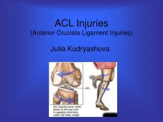
Cruciate Ligaments • Anterior Cruciate (ACL) • Prevents the tibia from moving anteriorly during weight bearing • Stabilizes the knee in extension and prevents hyperextension • Stabilizes against excessive internal rotation and serves as a secondary restraint to valgus/varus stress • Works in conjunction with hamstrings • Tight in extension and loosens during flexion (2 components) • Posterior Cruciate (PCL) • Taut throughout full ROM • Prevents excessive internal rotation
Menisci • Medial and lateral help to improve stability of knee, increase shock absorption and distribute weight over larger surface area • Move relative to knee flexion and extension in order to deal with contact forces • Patella • Aids in knee extension • Lengthens the lever arm of the quadriceps muscle • Distributes compressive forces on the femur by increasing contact area • Protects the patellar tendon against friction • Encounters gradual translation proximally and laterally during knee flexion (with associated internal rotation)
Muscle Function • For proper function the muscles surrounding the knee must work in concert with each other • Knee Flexion • Bicep femoris, semimembranosous, semitendinosous, gracilis, sartorius, gastrocnemius, popliteus and plantaris • Knee Extension • Rectus femoris, vastus lateralis, intermedius and medialis • External Rotation • Biceps femoris • Internal Rotation • Popliteus, semitendinosous, semimembranosous, sartorius, gracilis • Dynamic Lateral Stability • Iliotibial band
Rehabilitation Techniques • Strengthening • Open vs. Closed Kinetic Chain Exercises • OKC exercises tend to increase tibial translation which may be contraindicated • OKC exercises also increase rectus femoris activity • Increased shear forces and patellofemoral compressive forces with OKC exercises (at specific angles) • CKC exercises tend to generate more vasti musculature strengthening and joint compressive forces than OKC exercises • CKC exercises may be best utilized during dynamic stability and functional movement techniques • Must be aware of potential joint stresses at varying degrees of motion during strengthening
Range of Motion • While ROM is generally lost due to injury, early mobilization can reduce the histological changes that ligamentous tissue encounter (decrease water content and collagen-cross linkage) • Controlled motion to patient tolerance is critical • Pitfalls in ROM acquisition • Imperfect surgical techniques • Joint capsule/ligament contractures • Muscular resistance due to pain • Must determine cause of limitations and treat accordingly • Joint Mobilization Techniques • Must re-establish accessory motions of tibiofemoral, tibiofibular and patellofemoral joints to ensure appropriate physiological motion
Rehabilitation Techniques for Ligamentous and Meniscal Injuries Medial Collateral Ligament Sprain • Pathomechanics • Injury occurs either proximally (resulting in minimal laxity) or nearer the insertion (less stiffness and more laxity) • May have associated damage to medial meniscus • Graded injury • Injury Mechanism • Result of laterally applied valgus force to knee, occasionally in concert with rotational forces • Rarely a non-contact injury
Rehabilitation Concerns • Immobilization is generally very effective (with intact ACL) • For adequate healing • ligament fibers must remain in continuity • adequate stress to stimulate healing must be provided • protection from harmful stresses • Possible residual laxity due to stretch of ligament • Minimal effect on knee function • Symptomatic treatment with weight bearing as soon as possible
Rehabilitation Progression • Initially RICE and modalities to treat pain and inflammation • Crutch progression to full weight bearing as tolerated • Progress to no lag in extension and normal gait • Immobilization may be necessary for 1-2 weeks with grade 2 sprains • Early ROM and quad strengthening should be begin within the first 2 days post-injury with grade I condition • Quad sets, STLR’s, knee slides, riding a stationary bike • Grade 2 injuries may require 4-5 days for inflammation to subside
As pain continues to subside and ROM improves OKC exercises can be initiated • CKC exercises can also begin as tolerated • PNF, plyometric exercises and functional activities should also be incorporated gradually to enhance dynamic stability • Grade 3 sprains will require increased period of bracing (2-3 weeks, 0-45 degrees and 2-3 weeks 0-90 degrees)
Criteria for Return • Regained full ROM • Equal strength bilaterally • No tenderness • Successful completion of functional performance tests
Lateral Collateral Ligament Sprains • Pathomechanics • Isolated injuries are rare due to secondary stabilizers • Often the result of stress placed on lateral aspect of knee • Majority of injuries occur at proximal and distal attachments • May also be associated with injuries to ACL, PCL, posterolateral joint capsule, and peroneal nerve • Injuries graded based on physical examination
Injury Mechanism • Result of varus stress on knee • Rehabilitation Concerns • Determine extent of laxity • Grade 1 or 2 injuries degree of weight bearing • Grade 3 sprain will result in 4-6 weeks non-operative management • If rotational instability is associated with grade 3 injury surgical repair will be necessary • Rehabilitation Progression • Follows the same course as MCL injuries • In cases of surgical intervention, bracing with partial weight bearing will be required for 4-6 weeks, followed by a gradual progressive rehabilitation program return to activity = 6 months
Criteria for Return • Regained full ROM • Equal strength bilaterally • No tenderness • Successful completion of functional performance tests
Anterior Cruciate Ligament Sprain • Pathomechanics • Often occurs with cutting and jumping activities • Injury generally involves a mid-substance tear • ACL injuries are graded (1,2,3) • ACL deficient knees – generally accepted that torn ACL will not heal • Exhibits rotational instability that may lead to functional disability • Additionally, athlete may experience tears in meniscus and degenerative joint changes due to instability
Injury Mechanism • Non-contact injury twisting motion with foot planted and the athlete attempts to change direction • Results in deceleration, valgus stress and external rotation • Occasionally internal rotation is involved • Knee hyperextension with internal rotation can also become a factor • Tears resulting from a valgus force and disrupting the MCL, ACL and medial meniscus are termed the unhappy triad • Stop-jump landing phase of movement has been implicated in injury as well • Risk Factors • Shoe-surface interaction • Femoral intercondylar notch, ACL size, lower extremity alignment • Hormonal evidence • Mechanics of the lower extremity and trunk
ACL Injury Prevention • Multiple on-going studies • Establishment of pre-habilitation programs • Proprioceptive balance training in conjunction with weight training and jump-landing strategies • NC-LEIPP – program that focuses primarily on group jump-landing strategies • Rehabilitation Concerns • Conservative approach • Allow acute phase of injury to pass and follow up with aggressive rehabilitation • If desired or required level of stability is not attained surgical intervention will be necessary • Approach utilized with more sedentary individuals • Surgical intervention • Largely dependent on patient selection
Patient selection • Highly athletic • Individual is unwilling to change lifestyle • Rotational instability and giving way with daily activity • Additional structures are also involved • Recurrent effusion • Failure of rehabilitation following 6 month period • Surgery is necessary to prevent early joint degenerative changes • Partial tears • Immobilization and rehabilitation vs. Surgical intervention • Surgical repair options • Mid-substance repair suture with splinting (direct repair = results are not always ideal) • Extra-articular structure that lies outside capsule is moved to mechanically impact ACL function internally • Less expensive, aggressive approach with earlier return to activity • Not always recommended to high level athletes
Intrarticular repair structure inserted that will mimic function of the ACL • Bone-patellar grafts (human autograft/allograft) • Gracilis and semimembranosous autograft, Achilles allograft • Synthetic grafts – not as successful • Autografts = avascularity, resulting in decreased tissue strength • Allografts = disease transmission and tissue rejection, rehabilitation must be slightly less aggressive due to tissue changes • Rehabilitation Progression (non-operative) • Control pain and swelling through RICE, modalities and NSAID’s • Immobilization for protection and comfort with ambulation on crutches until extension is recovered (no extension lag in gait)
Progress immediately to quad set, STLR’s to regain motor control and prevent atrophy • Early ROM work as well (heel/wall slides, stationary bike) • OKC exercises • Flexion and extension exercises – restrict motion to 0-45 degrees for first 8-12 weeks • Gastroc and hamstring strength is critical • CKC exercises • Encourage co-contractions
Utilization of PNF patterns that stress tibial rotation • Functional knee brace utilization – controversial • Is it functional • Joint position sense feedback • Patient instructions with regard to activity • Lifestyle changes may be necessary to prevent further injury and the need for surgery • Surgical Intervention • Conservative vs. Accelerated • Slow progression emphasis • Slow progression to flexion and extension • Partial non-weight bearing post-operatively • CKC exercises at 3-4 weeks • Return to activity 6-9 months • Accelerated protocol • Immediate motion and weight bearing to tolerance • Early CKC for strength and neuromuscular control • Return to activity 2 months and competition 4-5 months
Pre-operative period (2-3 weeks) • Improve ROM, decrease pain and swelling • Re-establish quad control and normalize gait prior to surgery • Post operative period • Changes in tensile strength • Early tensile strength levels are high = aggressive rehab • Graft necrosis (6 weeks) revascularization (8-16 weeks) and remodeling (16 weeks on) • Swelling Control • RICE (Cryocuff) and modalities • Significant swelling will limit quad firing • Bracing • Locked in either full extension or 0-90 degrees passive and 40-90 degrees active ROM for first 2 weeks • Brace will be used until flexion exceeds limits of brace
Weight bearing (2-6 weeks) • Begin at 50% and work to full • Crutches can be removed with swelling is minimal, no extension lag and good quad strength with relatively normal gait • Range of motion • Begin immediately • Use of CPM units • Must regain knee extension • Active knee extension should be limited (60-90 degrees) • Active knee flexion should be achieved (90 degrees) by end of second week • Full flexion at weeks 5 or 6 • At 100-110, cycling can begin to help regain ROM
Early patellar mobilization instruction to enhance return of ROM • Strengthening • Avoid high level of stress on graft early on • Controlled strengthening of all muscles with emphasis on hamstrings • CKC exercises can begin when 90 degrees of flexion is achieved (1-2 weeks) • Minimize OKC exercises to reduce stress/shear forces
Isokinetic testing • Timing relative to rehabilitation program • PNF strengthening – rotational component is critical • Initiate progressive resistance at 5 months (OKC exercise) • Neuromuscular control • In addition to CKC exercises BAPS board (seated) should also be integrated • Balance training (BAPS board, Fitter) • Cardiorespiratory endurance • Cycling, UBE • Walking on treadmill can begin at 3 weeks • Swimming at 4-5 weeks, X-country skiing 6-7 weeks • Jogging and running 4-6 months depending on program • Functional training • Gradually incorporate running, jumping and pivoting activities in controlled environment • Single, double leg hop, carioca, shuttle runs, vertical jump • 5-6 weeks for accelerated program and 4 months for traditional training
Movement Technique Assessment • Determine deficits and incorporate into training progression • Assess technique and muscle activity • Eliminate areas of predisposition for future injury • Criteria for Return • Physicians will vary depending on surgical techniques and rehabilitation progressions • Vary anywhere from 4-12 months • No joint effusion • Full ROM • Isokinetic testing indicates quad and hamstring strength within 85-100% of uninvolved side • Satisfactory ligament stability (KT 1000) • Successful progression walking running • Successful performance during functional testing
Posterior Cruciate Ligament Sprains • Pathomechanics • Not commonly injured in athletics • Often concurrently injured with ACL, MCL, LCL or menisci • Controls rolling and gliding of tibia with ACL • Prevents posterior translation of the tibia on the femur • Meniscal lesions and chondral defects are likely with PCL deficient knees • 70% of tears occur at the tibia while the remaining 30% are split at the femur and mid-substance • Graded injury
Injury Mechanism • Knee is forced into hyperflexion with foot plantar flexed • Posteriorly driven tibia on fixed femur, or anteriorly forced femur on tibia • Knee hyperflexion with downward force on thigh • Hyperextension may result in combined PCL/ACL injury • Rehabilitation Concerns • Altered arthrokinematics • Surgical vs. non-operative approach
Rehabilitation Progression (non-operative) • Swelling and pain control initially • Immobilization for comfort and protection initially • Generally few limitations = quick progression • Early ROM and strengthening should be initiated • General strengthening • Focus on quadriceps • Work initially in the 20-45 degree range • Avoid OCK hamstring work due to posterior tibial translation • Incorporate CKC exercises to emphasize co-contractions • Functional knee braces are often not recommended • Possible proprioceptive/joint position sense benefits • Athlete should avoid repetitive stressful activities due to the incidents of progressive degenerative changes
Surgical Intervention • Maturation and healing process not well documented • Limit pain and swelling with RICE and NSAID’s • Immobilization in full extension for first week • During second week the brace should be unlocked for ambulation and PROM exercises • The brace should be worn for 4-6 weeks until flexion reaches 90-100 degrees • Crutch use should occur for 4-6 weeks until full weight bearing and can achieve full extension • Quad exercises and general hip musculature strengthening should begin at weeks 2-4 • Again, avoid knee flexion • At 4-6 weeks CKC exercises should begin • Utilize terminal knee extension
Incorporate neuromuscular control activities • Cycling can begin at 6 weeks with appropriate range (100-110 degrees) • Progress to jogging at 9 months • Gradually incorporate functional training • Criteria for Return • No joint effusion • Full ROM • Isokinetic testing indicates quad and hamstring strength within 85-100% of uninvolved side • Successful progression walking running • Successful performance during functional testing
Meniscal Injury • Pathomechanics • Aids in stability, acts as a secondary restraint in checking tibiofemoral motion, serves as shock absorber • Medial meniscus has higher incidence of injury • May be due to additional attachments via coronary ligaments – disruption due to valgus stress • Locking at 10-30 degrees is indicative of medial meniscus tearing while locking at 70 degrees is seen with posterior portion of lateral meniscal tear • May be longitudinal, oblique, transverse, vertical-longitudinal (bucket-handle tear)
Injury Mechanism • Weight bearing and rotation while flexing or extending knee • Valgus or varus force • Rehabilitation Concerns • Initially a conservative “wait and see” approach • Attempt to get through season and then perform surgery • Atrophy of edges will occur over time • Portions may become detached and wedged (chronic locking) • Recurrent swelling and decreased functional ability • Surgical intervention • Type of surgery • Location of lesion
Rehabilitation Progression (non-operative) • As signs and symptoms subside the athlete may return to activity • Minimize pain and inflammation • May require 3-5 days of limited activity prior to return • Partial menisectomy • Control pain and swelling via modalities and NSAID’s • Move to full weight bearing as soon as tolerated without limp or extension lag • Early pain free ROM and strengthening can begin with gradual addition of OKC and CKC exercises • Functional activities should be incorporated as the athlete feels ready (may occur within 3-6 days of surgery, more likely 2 weeks)
Meniscal Repair • Will involve absorbable sutures, vascular access channels and fibrin clot insertion • Complications are minimal if capsular damage is not present • Limit joint motion initially to allow for healing • Cardiovascular training (UBE) should be incorporated during immobilization • Lock in full extension for 2 weeks – allow partial weight bearing (full weight bearing after 6 weeks) • Quad sets along with hip abduction and adduction can be incorporated while in brace • Range is limited to 20-90 degrees for weeks 2-4 and 0-90 for weeks 4-6 • Gradually incorporate ROM exercises
At 6 weeks, brace can be removed • Rehabilitation should progress as previously discussed • Regain normal ROM and strength • Generally return to activity at about 3 months • If ACL injury is also involved, meniscal repair and healing constraints must be factored in to rehabilitation • Meniscal Transplant • Allograft or synthetic material • Lock in full extension for 6 weeks • Unlock only for PROM exercises from 0-90 degrees • Incorporate isometric quad sets and hip exercises • Partial weight bearing on crutches • Progress to full weight bearing with brace unlocked at 6 weeks discontinue use at 8 weeks or full extension, 100 degrees flexion and normal gait
Progressive strengthening, ROM and functional training can then begin • Full return is expected in 9-12 months • Criteria for Return • Swelling does not occur with activity • Full ROM has been regained • Equal bilateral strength in knee flexion and extension • Athlete can successfully complete functional progression
Rehabilitation for Patellofemoral and Extensor Mechanism Injuries Patellofemoral Stress Syndrome (PFSS) • Pathomechanics • Exhibit relatively common symptoms • Non-specific anterior knee pain, pain with stair climbing/descending • “Moviegoer’s sign” • Giving way of knee • Assess static alignment • Dynamic alignment • Stepping and bilateral/unilateral squats • Determine patellar tracking • Static and dynamic stabilizers must operate within a balance
Dynamic Stabilizers (continued) • Q-angle – may alter lateral valgus vector force (encourage lateral tracking) • A-angle – patella vs. tibial tubercle • Iliotibial band – causes lateral patellar tracking • Vastus medialis insufficiency – isotonically active throughout ROM, phasically active in individuals with patellofemoral pain and tends to lose fatigue resistant capacity • Vastus lateralis – tightness or muscle imbalance may cause lateral tracking
Excessive pronation – results in obligatory internal rotation altering mechanics at knee = lateral valgus vector force • Tight hamstrings – Results in increased knee flexion altering foot mechanics and presenting knee with additional lateral valgus vector force • Tight gastrocnemius – results in increased pronation • Patella alta – knee flexion occurs before patella is stabilized – lateral subluxation tendency increases • Patella baja – often results following injury or surgery • Patellar orientation • Glide component • Tilt component • Rotational component • Anteroposterior tilt
Rehabilitation Concerns • Avoid exacerbating activities • Improve lower limb mechanics and neuromuscular control • Enhance quad strength • Correct patellar positioning and tracking • Rehabilitation Progression • Strengthening • CKC exercises are recommended (reduced anterior shear) • Exercises that reduce compressive forces • Mini-squats, lateral step ups, stationary bike, slide board • Patella tracking and positioning • McConnell taping • Incorporates stretching (patellar glides) of lateral structures and patella positioning
Patellofemoral taping • Glide component – tape with knee in full extension, pull patella medially • Tilt component (positive lateral tilt) – tape in 30-40 degrees flexion, tape from middle of patella and pull medially • Rotational component (positive ER) – tape in 30-40 degrees flexion, pull inferior border upward and medially while rotating superior pole • Anteroposterior tilt – tape in full extension, tape over upper half of patella and press posteriorly with equal pressure • Reassess activity following taping procedure • Tape should be applied 24 hours daily initially (adjust as necessary)
Re-establishing Neuromuscular Control • Improve timing of VMO – isolate VMO function • VL:VMO ratio should be 1:1 • Utilization of biofeedback can be very useful in this muscle re-education • Must function concentrically and eccentrically throughout the ROM • Adduction exercises can be useful to facilitate VMO function due to the fact that VMO arises from adductor magnus • Mini-squats, step-ups/downs and leg presses for concentric/eccentric control, BAPS board for proprioceptive training
Criteria for Return • Taping should continue through training period • Maintain VMO activity for 5 minutes during walking gait • Can perform step ups for 1 minute with concomitant VL activity • Gradual weaning off process with tape • Tape can be left off completely when step ups can be performed for 5 minutes with no loss of VMO activity and half squat for 1 minute without VMO loss
Chondromalacia Patella • Pathomechanics and Injury Mechanism • Consequence of PFSS or from a direct impact to the patella • Softening or deterioration of the articular cartilage • Swelling and softening • Fissuring of softened cartilage • Deformation of the surface caused by fragmentation • No specific cause – generally associated with poor tracking (not always the case) • Pain in anterior region of knee with associated crepitus