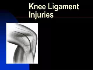
Evaluation of Common Knee Injuries
570 likes | 1.01k Vues
Evaluation of Common Knee Injuries. Frank J. Domino, M.D. Professor Family Medicine Clerkship Director Dept. Family Medicine & Community Health Un. Of Massachusetts Medical School Worcester, MA dominof@ummhc.org. Disclosure. Editor in Chief 5 Minute Clinical Consult

Evaluation of Common Knee Injuries
E N D
Presentation Transcript
Evaluation of Common Knee Injuries Frank J. Domino, M.D. Professor Family Medicine Clerkship Director Dept. Family Medicine & Community Health Un. Of Massachusetts Medical School Worcester, MA dominof@ummhc.org
Disclosure • Editor in Chief 5 Minute Clinical Consult • Author and Editor for Up To Date • Pri Med Curriculum Committee • Author/Editor: Familydoctor.org, Epocrates.com, Rxpalm, Inc.
Sports Medicine &Ambulatory Orthopedics • By the end of this session, you will: • Understand the anatomical and mechanical factors of knee injuries • Become familiar with the common presenting complaints • Learn to use appropriate history and physical exam skills to evaluate patients with common knee problems
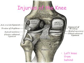
Knee Anatomy Right Knee
PE Knee – general points Valgus – Knock Knees Varus – Bow Legged Strain – injury to a muscle Sprain – injury to a ligament
Approach to Knee Examination I. History II. Observe: Effusion III. Palpation IV. ACL/PCL Evaluation V. Mensical Evaluation VI. Collateral Ligament Evaluation VII. Consider: ITB, Anserine Bursa, Patella, L4, Hip
I. History • Mechanism of Injury: • Timeline of pain and swelling • History of Trauma or injury • New Activities • Location of pain • Timing of Pain Onset: New Activities • Modifiers: WORSE- ↑ ↓ Stairs
II & III. Physical Exam Observe: Effusion Palpation ACL/PCL Injury (< 2 Hr)Meniscal Injury (> 2 Hr)
Chronic Effusion • DDX: Baker’s Cyst, DVT, Plica • Baker’s Cyst • Increase in Synovial Fluid • 2˚ Irritation • OA, Meniscus tear) • Plica: embroynic synovial remnant; Medial common
Case # 1 “I Felt a Pop” 22 yo female basketball player made a cut during practice and felt a “pop” in her knee with sharp pain. Significant knee swelling over 2 hours Over the last week the swelling has gone down She notes it feels like it “gives way”
What is your Differential Diagnosis? Anterior Cruciate Ligament Injury Meniscal Tear Patella Subluxation MCL Sprain Fracture
ACL Injury Foot planted, rotary injury Felt or Heard a Pop Swelling within first 2 hours Sense leg is lax + Ant. Draw or Lachman Evaluation: Plain Xrays MRI if Surgical Candidate Treatment: Effusion Drainage Repair: Transplant or Just Rehabilitation
Case #2. Lateral Knee Pain 44 year old male playing pick up basketball with some friends Doesn’t recall any injury, but next day noted some swelling and pain over the lateral knee “Hurts to go down stairs”
Lateral Knee Pain Differential Diagnosis: Lateral Meniscus Injury Iliotibial Band Syndrome Lateral Collateral Ligament Injury Bone Injury (Fracture, Contusion) Patello-Femoral Pain Syndrome (later)
DX: Meniscus Injury Rotary Injury Unilateral Pain May feel “Locking” Pain ↓ stairs + McMurry Eval: X Rays Tx: Rehab; Surgery if rehab fails or young Caveat: With edema comes atrophy of the VM
V. McMurray’s Test for Meniscal Injury Valgus Varus Medial Meniscus Lateral Meniscus • Medial Meniscus (Opposite foot rotation) • --Valgus stress & external rotation of foot • --Extend knee. • --Audible or palpable snap during extension + MM Tear • Lateral Meniscus • --Varus stress & internal rotation of foot • --Extend knee +LM Tear
VI. Collateral Testing • Hx: Clipping Injury • Patient Supine, Knee flexed at 10-20 degrees, • Apply Medial (vaRus ) force to test Lateral Collateral Ligament, • Apply Lateral (vaLgus) force to test Medial Collateral Ligament • (+) Pain or laxity
VI. Other: Iilotibial Band Syndrome Noble Compression Test Iliotibial Band Lateral View
Palpate Joint Line & Below:Lateral Knee Pain Ilio-tibial (Friction) Band Syndrome ??? Most common cause of Lat Knee pain in Runners Sloping road (Right in US) Insidious Onset with Running Evolves to pain with ADL Tx: Run Flat Surface Ice Massage, NSAIDs
Case 3: “Time to get in Shape” 53 year old male decides its time to get into shape; runs 3 miles first day out Presents with Medial Knee pain that seems worse at rest Does not recall any trauma or pain with the run
Medial Knee Pain Differential Diagnosis: Pes Anserine Bursitis Medial Meniscus Injury Medial Collateral Injury Tibial Head Fracture/Contusion Degenerative Joint Disease
Anserine Bursa • New Activity • Medial Pain • Pain at Rest • Tight Hamstrings • ? Related to DJD Medial View Attachment of insertion of tendon formed by Gracilis, Sartorius, & Semitendinosus Tendons
Pes Anserine Bursitis Diagnosis: History and PE Evaluation: No X Rays needed Treatment: NSAIDS, Ice Massage Rehab after acute injury Patient Education about new activities
Case #4: Hot Knee • 74 year old male presents with an acutely swollen, painful knee. No history of trauma, no new activities. • “Hurts all over” • Had too much Shrimp and wine at last night’s social
Case #4: Hot Knee Differential Diagnosis • Subclinical Fracture • Gout • Osteoarthritis • Infectious Arthritis • Ruptured Baker’s Cyst
Tap or No Tap If hot, or patient ill, refer. If you suspect Crystal Disease, and feel comfortable draining the knee using sterile technique, you may, BUT…
Case #5: 50 year old Male • No Recent Injury • Knee Swells by end of day • Very stiff and hard to walk when 1st OOB • Hx: recurrent trauma/surgeries
Osteoarthritis Medial > Lateral 6 commonly found Issues: 1. >50 years of age; 2. Morning stiffness < 30 minutes 3. Crepitus 4. Bony tenderness 5. Bony enlargement 6. No Erythema • X-rays (Wt Bearing): decrease joint space, osteophyte. • MRI: If considering surgery; Meniscal tears common; unclear if clinically relevant
Noises of the Knee “Crepitation”, “Clicking”, “Grinding”, Popping, and “Snapping” sounds. Associated with: osteoarthritis, patellofemoral syndrome, meniscal tears and iliotibial band syndrome.
Osteoarthritis: Treatment 1. Analgesic: Acetaminophen 1 Gm QID, NSAIDs & GI protection… Glucosamine? 2. Rehab: PT, Non-Wt Exercise, Wedge Insert Water Aerobics; Appl Nurs Res. 2010 Dec 28. 3. Weight Loss 4. Steroid Injection ??? 5. Off Loading Brace 6. Invasive/Arthroscopic/ Replacement Jordan KM; Ann Rheum Dis 2003 Dec;62(12):1145-55
Immobilizer Unloading OA Brace A/P/Lat Stabilizing
One CM Above One CM Lateral To Superior, Lateral Pole of Patella Angle Needle 45 Degrees distally & Down
Case: #6. 15 y/o Female Volleyball Player • No History of Trauma or acute pain • Insidious Onset Generalized pain • Now Pain w/ Flexion of Knee • First, only with sports, now with walking, going up and down stairs
Generalized Knee/Patellar Pain 1. Patello-Femoral Syndrome • Proximal/Generalized Patellar Pain • 2° Quadriceps Dysfunction • Tight ITB & Weak VMO: Patella track laterally and compresses into the lateral femoral condyle. 2. Jumper’s Knee • Distal Patellar Pain • Distal Patellar Tendon Inflammation 2° Tight Hamstring and Quadriceps
Patellar Tendon Anatomy All One Tendon PF Syndrome
Dx: Anterior & Generalized Knee Pain • Patello-Femoral Syndrome: • Patellar Grind Test: applying gentle downward pressure w/leg extended & VMO CTX • Lateral Patellar Glide Test: laterally directed pressure to patella of extended leg & VMO CTX -Contraction of quadriceps and/or pain implies VMO-IT discrepancy • Jumper’s Knee: • Downward pressure to Proximal Patella with palpation under Distal Patella -Pain or apprehension
Tx: Generalized Knee Pain • Patello-Femoral Syndrome: • RICE, Physical Tx. Neoprene Sleeve or Subpatellar strap w/activities • Jumper’s Knee (Patellar Tendonitis): • RICE, Physical Therapy, Stretching (esp. of Hamstrings) & Massage
Imaging: Ottawa Knee Rules • TRAUMA or Suspicion—Films! • 96% Sensitive; ~50% Specific • Age 50(55) years • Patellar tenderness • Fibular Head Tenderness • Inability to flex the knee to 90 degrees • Inability to bear weight both immediately & in the E.D. for 4 steps, regardless of limp
References • www.arthritispractitioner.com/article/5891 • www.kneeclinic.info/knee_problems.php • www.nismat.org/orthocor/ exam/knee_eval.html • http://www.sportsdoc.umn.edu/ • www.tcusportsmedicine.com • www.aafp.org/fpm/20050300/67atoo.html • www.hughston.com/hha/a.extmech.htm • www.emedicine.com/sports/topic56.htm
Questions? Frank.domino@umassmemorial.org