Sensory Physiology
480 likes | 530 Vues
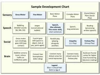
Dive into sensory physiology to understand how stimuli are sensed, processed, and integrated by the body's pathways, from receptors to the cerebral cortex and brain stem. Learn about somatosensory receptors, stimulus properties, sensory coding, and more.

Sensory Physiology
E N D
Presentation Transcript
Sensory Pathways and Integration • Stimulus as physical energy sensory receptor • Receptor acts as a transducer • Intracellular signal usually change in membrane potential • Stimulus > threshold action potential to CNS • Integration in CNS cerebral cortex or acted on subconsciously • Sensory information travels either from the spinal cord to brain by ascending pathway or directly to brain stem via cranial nerves • Visceral reflexes are integrated in brain stem or spinal cord and usually do not reach conscious perception. For example- pupil dilation • Perceptual threshold is the level of stimulus necessary to be aware of particular sensation
Somatosensory Receptors Figure 10-1a
Sensory Neurons: Two-Point Discrimination The more regions that overlap, the lesser the sensitivity for point discrimination. Why not have the same degree of discrimination everywhere? Figure 10-3a
Properties of Stimulus: Location Without receptive fields, the brain uses timing of receptor stimulation for sound. A sound created to the side of a person will not reach receptors on both sides simultaneously, thus allowing for localization of sound A sound created in front of a person reaches both receptors simultaneously. Figure 10-5
Properties of Stimulus: Location Lateral inhibition creates more contrast between activated receptive fields and inactive neighbors – activated receptor sends inhibiting signals to neighboring receptors. Figure 10-6
Properties of Stimulus • Intensity- the strength of a stimulus • Coded by number of receptors activated (population coding) and frequency of action potentials (frequency coding) • Duration- for how long a stimulus is present • Coded by duration of action potentials – the primary sensory neuron will produce action potentials as long as a stimulus ispresent. • Some receptors can adapt or cease to respond- adaptation allows the brain to get rid of stimulus info that does not threaten homeostasis • Tonic receptors versus phasic receptors • Tonic receptors- fire rapidly when activated yet slowly adapt to a stimulus but continue to signal as long as the stimulus remains at the same intensity. • Phasic receptors- fire when activated but are quick to adapt to the stimulus as long as it is of continuous strength.
Properties of Stimulus (a) Stimulus 20 0 Amplitude Membrane potential (mV) -20 -40 Threshold -60 -80 Duration 0 5 10 0 5 10 0 5 10 Time (sec) (b) Longer and stronger stimulus 20 0 -20 Membrane potential (mV) -40 -60 -80 0 5 10 A stronger stimulus will signal well above threshold and for a longer time thus creating more action potentials. Figure 10-7 (4 of 6)
Properties of Stimulus (a) Stimulus 20 0 Amplitude Membrane potential (mV) -20 -40 Threshold -60 -80 Duration 0 5 10 0 5 10 0 5 10 Time (sec) (b) Longer and stronger stimulus 20 0 -20 Membrane potential (mV) -40 Threshold -60 -80 0 5 10 0 5 10 0 5 10 Sensory coding for stimulus intensity and duration As long as the stimulus goes beyond threshold action potentials will fire. They will continue to fire proportional to intensity and duration. Frequency of action protentials is proportional to stimulus intensity and to release of neurotransmitters Figure 10-7
Tonic and Phasic Receptors Tonic receptors fire rapidly when activated yet slowly adapt to a stimulus but continue to signal as long as the stimulus remains at the same intensity. Figure 10-8a
Tonic and Phasic Receptors Phasic receptors fire when activated but are quick to adapt to the stimulus as long as it is of continuous strength. Figure 10-8b
Somatic Senses: Modalities & Pathways • Touch- fine, pressure, deep pressure, vibration, texture, ect. • Proprioception- position of joints/muscle, stretching • Temperature- free nerve endings that determine hot/cold touch, more cold receptors, do not adapt if tissue damage is possible. • Nociception • Pain- may prevent injury or signal site of injury • Itch- a type of pain receptor that can be activated by the release of histamine.
The Somatosensory Cortex Figure 10-10
Temperature Receptors • Free nerve endings • Terminate in subcutaneous layers • Slow to adapt between 20oC-40oC, do not adapt where tissue is likely to get damaged • Cold receptors • Respond to temperatures lower than body temperature • More abundant than warm receptors • Warm receptors • Respond to temperatures above body temperature to about 45°C • Pain receptors activated above 45°C
Nociceptors • Free nerve ending • Respond to strong noxious stimulus that may damage tissue • Pain is a perception not a stimulus • Modulated by local chemicals • Substance P is secreted by primary sensory neurons • Mediate inflammatory response • Inflammatory pain • Reflexive protective response • Integrated in spinal cord • Withdrawal reflex • Ascending pathway to cerebral cortex • Becomes conscious sensation (pain or itch)
Nociceptors: Pain and Itch • Itch: Histamine activates C fibers causing itch • Pain • Subjective perception – Not a true stimulus but an interpretation by the brain. If signals do not reach the brain pain is not perceived even though there may be damage. • Fast pain- initial sensation, draws attention to area • Sharp and localized—by A fibers • Slow pain – secondary sensation, may seem dull, can become chronic • More diffuse—by C fibers
The Gate-Control Theory of Pain Figure 10-12a
Pain: Referred Pain Sharing a secondary neuron converges the signals into a single tract, thus it is more difficult to distinguish it from the somatic areas Figure 10-13b
Pain • Ischemia • Lack of adequate blood flow – tissue damage will initiate a pain signal which can also be interpreted as referred pain • Example- heart muscle deprivation of oxygen will cause referred pain over chest, face, and arm • Chronic pain is a pathological pain- also called neuropathy and not fully understood. • Analgesic drugs • Aspirin • Inhibits prostaglandins and slows transmission of pain to site of injury • Opiates like morphine (natural version= endorphins) • Act on the CNS opoid receptors blocking NT release of primary sensory neurons or postysnaptic inhibition of secondary sensory neurons
Olfaction • Link between smell, memory, and emotion • Vomeronasal organ (VNO) in rodents • Response to sex pheromones • Olfactory cells • Olfactory epithelium in nasal cavity • Odorants bind to odorant receptors, G-protein-cAMP-linked membrane receptors
Anatomy Summary: The Olfactory System Figure 10-14a
Summary of Taste Transduction Figure 10-16
Summary of Taste Transduction Each taste bud has different receptors that can be stimulated by a specific molecule. Each receptor causes a different intracellular reaction that is interpreted as a certain taste Humans and animals may develop specific hungersuch as salt appetite Figure 10-16
Anatomy Summary: The Ear The ear is a sense organ that is specialized for two functions: hearing and equilibrium Figure 10-17
Sound Waves • Hearing is our perception of energy carried by sound waves. • Frequency is translated into pitch • Intensity is translated as loudness Figure 10-18a
Sound Transmission Through the Ear Figure 10-19 (6 of 6)
Anatomy Summary: The Cochlea • Perilymph in vestibular and tympanic duct • is similar to plasma • Endolymph in cochlear duct • is secreted by epithelial cells • and is similar to intracellular fluid
Anatomy Summary: Hair cells of the Cochlea • Hair cell of the choclea surrounded by endolymph • Repeated exposure to loud sounds cause hair cell death that eventually leads to hearing loss.
Signal Transduction in Hair Cells The apical hair cell is modified into stereocilia The change in direction opens or closes ion channels changing ultimately affecting the amount of action potentials Figure 10-21a
The Ear: Auditory Pathways and hearing loss • Air waves cause vibration which causes fluid movement that is translated into electrical signals in cochlea • Electrical signals travel from primary sensory neurons to nuclei in medulla oblongata via cranial nerve VIII • Signal travel from medulla to midbrain and thalamus • Ipsilateral and contralateral signals arrive to auditory cortex of each cerebral hemisphere • Conductive Hearing Loss • No transmission through either external or middle ear • Central Hearing Loss • Damage to neural pathway between ear and cerebral cortex or to cortex itself • Sensorineural Hearing Loss • Damage to structures of inner ear
Anatomy Summary: The Vestibular Apparatus Figure 10-23a
Anatomy Summary: The Vestibular Apparatus Figure 10-23b
Rotational Forces in the Cristae The semicircular canals sense rotational acceleration Figure 10-24
Otolith Organs The otolith organs sense linear acceleration and head position Figure 10-25a
The Ear: Equilibrium Central nervous system pathways for equilibrium Figure 10-26
The Eye and Vision • Light enters the eye through the pupil • Size of the pupil modulates light • Photoreceptors transduce light energy into electrical signals sent through neural pathways and processed at visual cortex • Shape of lens focuses the light on retina • Pupillary reflex • Standard part of neurological examination • The iris is the pigmented part of the eye, the muscle of the iris contract to change the size of the pupil
External Anatomy of the Eye Figure 10-27
Anatomy Summary: The Eye Figure 10-28a
Anatomy Summary: The Eye Figure 10-28b
Refraction of Light Figure 10-30a
Mechanism of Accommodation Accommodation is the process by which the eye adjusts the shape of the lens to keep objects in focus Figure 10-32a
Anatomy Summary: The Retina Light bounces off the pigmented epithelium to activate the receptors Figure 10-35d
Vision: Phototransduction Rods and cones only respond to photons within the visible ligth range. Image projection onto the retina causes the image to be flipped upside down Figure 10-36
Vision: Photoreceptors Rods and cones Color-blindness results from a defect in one or more of the three types of cones Figure 10-37
Vision: Neural Pathways for Vision and the Pupillary Reflex Figure 10-29a
Vision: Neural Pathways for Vision and the Pupillary Reflex Figure 10-29c
Visual Fields and Binocular Vision Items that fall within the binocular zone are perceived in 3D and allow for depth perception. Figure 10-41