Download
1 / 19
190 likes | 289 Vues
This case report discusses the cardiovascular disorder in a 78-year-old ex-smoker with aortic stenosis, focusing on diagnosis, clinical presentation, and treatment options.

E N D
Casereport no. 1,Department Pathological Physiology V. Danzig, MD, PhD,2nd Dept. Internal MedicineCardiology andAngiologyDivision 1st Med.F CUNI
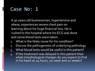
Patient history • 78 yr. old male, retiree, worked as mechanical engineer • Family history: longevity, cardiovascular complications, yet no early deaths • (Ab)usus: quitted smoking 15 yrs ago, 10-15 cigarettes daily before, alcohol drinking denies • Patient history: repeated herniotomy, benign prostatic hypetrophy, diabetes mellitus last 6 years – on diet
Patient history- questions • What is the risk (high/ medium/ low) of cardiovascular disease in this 78 year old former smoker? • When is the onset of cardiovascular disorder considered early and when timely? • What are the effects of smoking on the cardiovascular system and on the respiratory system (related to malignant tumors), what are the differences?
Current disorder • Last 3 years progression ofexertional dyspnea and chest pain projecting to medium and lower sternum • Progressive worsening last 2 weeks, pains in rest • Once a pain attack in walking, loss of consciousness, awakened lying on ground • Admitted to hospital after a nigh long lasting retro-sternal pains, dyspnea, felt forced to sit, first time felt as well irregular and fast heardbeats
Current disorder- questions • Give clinical definition of dyspnea. What is its origin in (left) heart disorder? Other causes besides cardiomyopathy? • Name/ description: ischemic myocardial pain, its mechanism/ cause? • Name/ description: of a short time loss of consciousness • What is the prime cause of myocardial ischemia in Czech population? What is differential diagnostics? • Synonyms for palpitations? They are signs of what diseases? What does it mean when patient complains that they are irregular?
What investigation methods to use in this patient • ECG: - what are signs of acutemyocardial infarction?Elevations of the ST segment? - cardiac rhythm ? • Biochemical markers – in this patient were not elevated – no myocardial necrosis • X-ray of heart and lungs – venostasis (oedema)
Definition of atrial fibrillation • Atrial fibrillation isan irregular atrial activity. ECG record shows no P waves. Instead there are fast oscillations, also denoted as “F” waves. Ventricular response are typically QRS complexes with RR intervals of different length.
Investigation methods - questions • How many leads are in standard ECG? Names and groups of leads? How we record continuous ECG and how many leads in it we use? • Name biochemical cardiac markers, describe them, what is their use? • What are signs of atrial fibrillation? What are the risks?
Further investigations • echokardiography, ECHO: - Findings: calcified aortal valve with limited cusp excursions, Doppler – tight stenosis w. gradients 65/ 34 mm Hg, mouth area 0.4 cm2/m2 (per body surface) - Ascendent aorta dilation - dilatace ascendentní aorty - EF (ejection fraction) LV 0,45 – 0,49 (= 45-49%) • selectivecoronarography: no pathology/ normal findings
Further investigations - questions • What gradient is important here (related to aortal valve and its stenosis) • What are two basic approaches to measure the gradient? Compare the two? Can we always use both of them? • What can be the complications? • Why we calculate the area? Explain cases when area is significantly reduced yet the high gradient is not present. • What is left ventricle ejection fraction, its units and normal values?
Therapy • Temporary therapy: aim to compensate – therapy of heart failure and control of ventricular response to atrial fibrillation • Definitive therapy: replacement of aortal valve by bio-prosthesis, eventually a plastic surgery of ascendent aorta
Therapy - questions • What is the relation of atrial fibrillation to the principal heart disorder (aortal stenosis)? • Why as a first step in the therapy of atrial fibrillation we need a slow down of the ventricular frequency? • Why and when a plastic surgery of aorta is needed as well?
Conclusions I • Aortal stenosis: is frequent and becoming yet more frequent acquired valve disorder, as the population is ageing. • This condition is followed up by a pressure overload of left ventricle. This propagates backwards and leads to atrial dilation (closing a vicious circle this way) and back to pulmonary circulation. • Typical compensatory mechanism is left ventricle hypertrophy. This is followed up by development of ventricle dilation, deterioration of systolic function and failure.
Conclusions II • Symptomes and their patho-physiological explanations (Q/+A): • dyspnoe • by back-propagationof elevatedintra-ventricular pressure • stenocardia • loweredthroughput in coronary arteries, esp. in tachycardia • exertional syncope • inability to elevatecardiac outputwhen valve mouth area is reduced • low pressure amplitude • slower onsetof systolic pressure in aorta • Therapy: aortal valve replacement (bio-prosthesis) by cardio-surgery. Transcatheter Aortic Valve Implantation in selected high risk patients.