Download

1 / 61
610 likes | 766 Vues
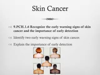
I am giving you guys skin cancer. RODENT ULCER MARJOLIN’S ULCER EPITHELIOMA. RODENT ULCER. What is it??. Usually a slow-growing , locally invasive malignant tumor of pleuripotent epithelial cells Arising from basal epidermis and hair follicles, hence affecting the pilosebaceous skin.

E N D
What is it?? • Usually a slow-growing, locally invasivemalignant tumor of pleuripotent epithelial cells • Arising from basal epidermis and hair follicles, hence affecting the pilosebaceous skin
Predisposing Factors • Exposure to UVR • Exposure to Arsenic compounds, coal tar, aromatic compounds, IR, Coal Tar • Genetic Skin Cancers • White skinned people • Age 40-80 yrs
PATHOGENESIS • No apparent precursor lesion • Proportional to initial dose of carcinogen but not duration. • Rarely metastise,Hard to culture,Resist transplantation • Mesodermal factors acting as intrinsic promoters coupled with an initiation step
MACROSCOPIC GENERALISED • LOCALISED • Nodular • Nodulocystic • Cystic • Pigmented • Maevoid 90% • INFILTRATIVE • Ice pick • Morphoeic • Cicatrising • SUPERFICIAL • Multifocal • Superficial Spreading
MICROSCOPIC Ovoid cells in nests with single outer Palisading layer
ORIGIN • Basal layer of epidermis. • Occasionally arises from basal cells of hair follicles and sweat glands. • Seen in scalp known as TURBAN TUMOR
Why Rodent Ulcer?? • LOCAL INVASION • Gradually destroys tissues it comes in contact with!! • LYMPHATIC SPREAD not seen • Regional lymph nodes NOT enlarged. • Blood spread rare
CLINICAL FEATURES • SYMPTOMS: • Persisting ulcer or nodule • Not painful / may itch • Grows slowly • SIGNS: • Site- 90% BCC seen on face above line joining angle of mouth to lobule of ear.
SIGNS: Site- 90% BCC seen on face above line joining angle of mouth to lobule of ear.
COMMON SITES • INNER CANTHUS OF EYE • OUTER CANTHUS OF EYE • NOSE • ON AND AROUND NASOLABIAL FOLD • ON THE FORE HEAD Tear Cancer
LESION • Starts as Nodule • Gradually centre of nodule dies and ulcer results. • EDGE- Raised & Rounded.BEADED MARGIN • GROWTH SPREADS- Shape irregular. • FLOOR- Dried Serum, Epithelial cells. • BASE- Tissue & Tumor is eroding ie. Fat, Muscle, Bone!!
PROGNOSIS LOW RISK • HIGH RISK • > 2cm • Eye, Nose , Ear • Ill defined margins • Recurrent ulcer
MANAGEMENT • SURGICAL • Excision • Mohs Micrographic Surgery • Two stage surgery • NON SURGICAL • Curettage • Electrodessication • Laser Vaporization RADIOTHERAPY • No pathological specimen • No confirmation of diagnosis • Tumor margin not confirmed
Indications • POORLY DEMARCATED • RECURRENT • INCOMPLETELY EXCISED • AREA AROUND EYES NOSE EAR
PROCEDURE • Performed under LOCAL ANESTHESIA • Initial SAUCERISING EXCISION of primary tumors visible extent. • Sample and the defect are then marked and oriented • Map of specimen drawn & characterised using different colored stains in different equators.
Histotechnician receives tissue sample from the Mohs Surgeon. • Sample is sectioned and stained with H&E • Mohs surgeon examines slide for tumor residue and excises the relevant mapped parts.
NON SURGICAL • Radiotherapy (scars badly) • Cryotherapy • Topical Chemo (5-fluorouracil, imiquimod)
Follow Up • Gross Margin involvement: 67% recurrence • Microscopic involvement: 33% recurrence within 2 yrs. • Uncomplicated completely excised: Surveillance as in HIGH Risk groups! • GORLIN’s syndrome
What is it?? • SCC is a malignant tumor of keratinising cells of the epidermis or its appendages. • Arises from the stratum basale of the epidermis • 2nd most common skin tumor (4 times less than BCC & affects Elderly)
PREDISPOSING FACTORS • WHITE SKINNED • TWICE AS COMMON IN MEN • SUN EXPOSURE • CLOSER TO EQUATOR
Contd. • chronic inflammation (chronic sinus tracts, pre-existing scars, osteomyelitis, burns, vaccination points) • immunosuppression (organ-transplant recipients). • When a SCC appears in a scar it is known as a Marjolin’s ulcer.
Contd • Radiation exposure • Smoking • HPV infection
MACROSCOPIC • The early appearance of SCC may vary from smooth nodular to verrucous, papillomatous and ulcerating lesions. • Eventually all lesions ulcerate
MICROSCOPIC • Solid column of epithelial cells that are seen growing down into dermis. • Expanding into bulb like masses. • KERATINISATION, CELL NEST/Epithelial pearl appearance.
SPREAD • LOCAL SPREAD • LYMPHATIC SPREAD • BLOOD SPREAD- rarely
HISTOPATHOLOGY • Pathological pattern (e.g. adenoid), • Cellular morphology (e.g. spindle) • Broder’s grade(grade 1 to 4) • Depth of invasion
ORIGIN • a) skin denovo • b)pre existing condition like • Long standing ulcers • Senile keratosis • Leukoplakia • Skin exposed to radiation • lupus
PREMALIGNANT LESIONS • Chronic Ulcer : MARJOLINS ulcer
FEATURES • Painless!!! • Less malignant than typical SCC • Edge not always raised & everted • Slow growing malignant lesion • No lymphatic metastasis
TREATMENT • Surgery : Wide excision of lesion with 1 cm margin.
OTHER PREMALIGNANT COND. Xeroderma pigmentosum • Bowen disease Senile keratosis
Condition which cause chr irritation • 1)Leukoplakia • 2)Burn,scar,venous ulcer,OM, • 3)Continous heat by a charcoal burner ie.kangri – abdomen -KANGRI CANCER
Prolonged exposure to chemicals as in soot – SCC of scrotum – CHIMNEY SWEEP CANCER
SITES SKIN- anywhere
CLINICAL FEATURES • History • Age > 40 yrs • Occupation -chimney sweepers • Duration- one or few months (variable cap growth) • Symptoms • Nodule/ Ulcer • Usually Painless • Enlarged Lymph node ( unlike BCC)
LOCAL EXAMINATION • Site • Size and shape.- circular /oval • EDGE: Raised & Everted • FLOOR: Necrotic tissue, Serum, Blood. • BASE: Indurated. • MOBILITY: early cases can be moved later fixed • Regional Lymph nodes enlarged due to 2ndry infectn • Tenderness +