Download
1 / 4
40 likes | 58 Vues
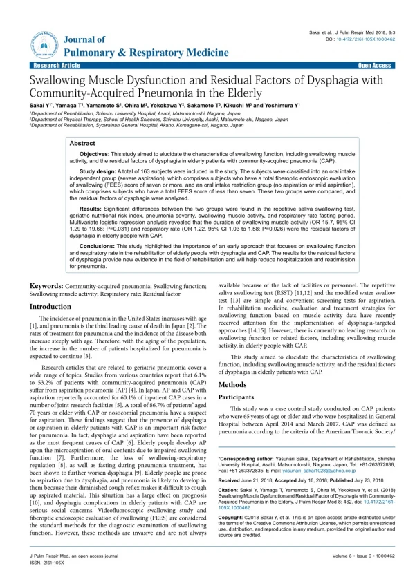
The incidence of pneumonia in the United States increases with age, and pneumonia is the third leading cause of death in Japan. The rates of treatment for pneumonia and the incidence of the disease both increase steeply with age. Therefore, with the aging of the population, the increase in the number of patients hospitalized for pneumonia is expected to continue.

E N D
Sakai et al., J Pulm Respir Med 2018, 8:3 DOI: 10.4172/2161-105X.1000462 JournalofPulmonary&RespiratoryMedicine ISSN: 2161-105X Journal of Pulmonary & Respiratory Medicine Research Article Swallowing Muscle Dysfunction and Residual Factors of Dysphagia with Community-Acquired Pneumonia in the Elderly Sakai Y1*, Yamaga T1, Yamamoto S1, Ohira M2, Yokokawa Y2, Sakamoto T3, Kikuchi M3 and Yoshimura Y1 Open Access 1Department of Rehabilitation, Shinshu University Hospital, Asahi, Matsumoto-shi, Nagano, Japan 2Department of Physical Therapy, School of Health Sciences, Shinshu University, Asahi, Matsumoto-shi, Nagano, Japan 3Department of Rehabilitation, Syowainan General Hospital, Akaho, Komagane-shi, Nagano, Japan Abstract Objectives: This study aimed to elucidate the characteristics of swallowing function, including swallowing muscle activity, and the residual factors of dysphagia in elderly patients with community-acquired pneumonia (CAP). Study design: A total of 163 subjects were included in the study. The subjects were classified into an oral intake independent group (severe aspiration), which comprises subjects who have a total fiberoptic endoscopic evaluation of swallowing (FEES) score of seven or more, and an oral intake restriction group (no aspiration or mild aspiration), which comprises subjects who have a total FEES score of less than seven. These two groups were compared, and the residual factors of dysphagia were analyzed. Results: Significant differences between the two groups were found in the repetitive saliva swallowing test, geriatric nutritional risk index, pneumonia severity, swallowing muscle activity, and respiratory rate fasting period. Multivariate logistic regression analysis revealed that the duration of swallowing muscle activity (OR 15.7, 95% CI 1.29 to 19.66; P=0.031) and respiratory rate (OR 1.22, 95% CI 1.03 to 1.58; P=0.026) were the residual factors of dysphagia in elderly people with CAP. Conclusions: This study highlighted the importance of an early approach that focuses on swallowing function and respiratory rate in the rehabilitation of elderly people with dysphagia and CAP. The results for the residual factors of dysphagia provide new evidence in the field of rehabilitation and will help reduce hospitalization and readmission for pneumonia. Keywords: Community-acquired pneumonia; Swallowing function; Swallowing muscle activity; Respiratory rate; Residual factor Introduction The incidence of pneumonia in the United States increases with age [1], and pneumonia is the third leading cause of death in Japan [2]. The rates of treatment for pneumonia and the incidence of the disease both increase steeply with age. Therefore, with the aging of the population, the increase in the number of patients hospitalized for pneumonia is expected to continue [3]. Research articles that are related to geriatric pneumonia cover a wide range of topics. Studies from various countries report that 6.1% to 53.2% of patients with community-acquired pneumonia (CAP) suffer from aspiration pneumonia (AP) [4]. In Japan, AP and CAP with aspiration reportedly accounted for 60.1% of inpatient CAP cases in a number of joint research facilities [5]. A total of 86.7% of patients’ aged 70 years or older with CAP or nosocomial pneumonia have a suspect for aspiration. These findings suggest that the presence of dysphagia or aspiration in elderly patients with CAP is an important risk factor for pneumonia. In fact, dysphagia and aspiration have been reported as the most frequent causes of CAP [6]. Elderly people develop AP upon the microaspiration of oral contents due to impaired swallowing function [7]. Furthermore, the loss of swallowing-respiratory regulation [8], as well as fasting during pneumonia treatment, has been shown to further worsen dysphagia [9]. Elderly people are prone to aspiration due to dysphagia, and pneumonia is likely to develop in them because their diminished cough reflex makes it difficult to cough up aspirated material. This situation has a large effect on prognosis [10], and dysphagia complications in elderly patients with CAP are serious social concerns. Videofluoroscopic swallowing study and fiberoptic endoscopic evaluation of swallowing (FEES) are considered the standard methods for the diagnostic examination of swallowing function. However, these methods are invasive and are not always available because of the lack of facilities or personnel. The repetitive saliva swallowing test (RSST) [11,12] and the modified water swallow test [13] are simple and convenient screening tests for aspiration. In rehabilitation medicine, evaluation and treatment strategies for swallowing function based on muscle activity data have recently received attention for the implementation of dysphagia-targeted approaches [14,15]. However, there is currently no leading research on swallowing function or related factors, including swallowing muscle activity, in elderly people with CAP. This study aimed to elucidate the characteristics of swallowing function, including swallowing muscle activity, and the residual factors of dysphagia in elderly patients with CAP. Methods Participants This study was a case control study conducted on CAP patients who were 65 years of age or older and who were hospitalized in General Hospital between April 2014 and March 2017. CAP was defined as pneumonia according to the criteria of the American Thoracic Society/ *Corresponding author: Yasunari Sakai, Department of Rehabilitation, Shinshu University Hospital, Asahi, Matsumoto-shi, Nagano, Japan, Tel: +81-263372836, Fax: +81 263372835; E-mail: yasunari_sakai1028@yahoo.co.jp Received June 21, 2018; Accepted July 16, 2018; Published July 23, 2018 Citation: Sakai Y, Yamaga T, Yamamoto S, Ohira M, Yokokawa Y, et al. (2018) Swallowing Muscle Dysfunction and Residual Factor of Dysphagia with Community- Acquired Pneumonia in the Elderly. J Pulm Respir Med 8: 462. doi: 10.4172/2161- 105X.1000462 Copyright: ©2018 Sakai Y, et al. This is an open-access article distributed under the terms of the Creative Commons Attribution License, which permits unrestricted use, distribution, and reproduction in any medium, provided the original author and source are credited. J Pulm Respir Med, an open access journal ISSN: 2161-105X Volume 8 • Issue 3 • 1000462
Citation: Sakai Y, Yamaga T, Yamamoto S, Ohira M, Yokokawa Y, et al. (2018) Swallowing Muscle Dysfunction and Residual Factor of Dysphagia with Community-Acquired Pneumonia in the Elderly. J Pulm Respir Med 8: 462. doi: 10.4172/2161-105X.1000462 Page 2 of 4 Infectious Diseases Society of America [16]. The exclusion criteria included the following: a history of central nervous system disorder, neck deformity, or cognitive decline (>23 points on the Mini-Mental State Examination) or lung disease such as interstitial pneumonia or chronic obstructive pulmonary disease. For each observation and test item, data collection was performed at the start of oral intake by CAP patients (they started direct therapy using jerry within approximately seven days after hospitalization). To mitigate the risk of aspiration, oral intake was initiated after the breaking of fever and the peaking of inflammation. The study protocol was approved by the Ethics Committee of our institution (No. 2014-4), and we obtained written informed consent from all participants after the study protocol was explained in detail. This study was conducted in accordance with the standards of the latest revision of the Declaration of Helsinki. Evaluation Data collection: We collected data on the age, sex, respiratory rate (RR), CURB-65 Severity Score, and fasting period of the patients. Laboratory data were collected for C-reactive protein (CRP), white blood cell (WBC) count, and blood urea nitrogen (BUN). Nutritional status was evaluated by the geriatric nutritional risk index (GNRI) and was calculated as follows: (1.489 × serum albumin level, g/dl)+(41.7 × current body weight/ideal body weight). Furthermore, the RSST, FEES, and muscle activity during swallowing were obtained by surface electromyogram (sEMG). The RSST was conducted according to published protocols [11,12]. The first and second finger pads were placed gently on the laryngeal prominence. Thereafter, the patient was asked to swallow saliva, and the evaluator confirmed laryngeal elevation as the swallowing reflex. The evaluator noted the point at which the laryngeal prominence passed the finger pad and moved further downward before returning to the original position; this was considered one swallow. The number of voluntary swallows within 30 seconds was counted. The basic protocols for FEES were followed [17]. FEES is a swallowing function test that is performed using a fiberscope or an electronic endoscope. The fiberscope or endoscope is passed just inferior to the inferior turbinate in the inferior meatus. When the scope is in the nasopharynx, the velopharyngeal port can be viewed adequately. The patient is requested to dry swallow to allow the assessment of velopharyngeal competence during swallowing. The tip of the scope is then deflected downward, and the scope is passed into the oropharynx. Finally, the scope is passed to a point posterior to the epiglottis, where the general appearance of the laryngeal structures is visualized. EMG observation and analysis methods: sEMG recording was performed using sEMG (Electromyograph MQ-Air, KISSEICOMTEC Co.). Recording electrodes are affixed to the suprahyoid and infrahyoid muscle groups according to previously described methods [18]. The subject assumes an upright sitting posture with the chin in the Frankfurt plane parallel position. Jelly is then used to confirm the presence of normal swallowing sound [19] in conjunction with simultaneous cervical auscultation by using a stethoscope. The consistency of the jelly (700 N/m2 hardness, 300 J/m3 adhesiveness, and room temperature) is the same as that of the initially swallowed food. Recordings from sEMG were full-wave rectified to yield wave rectification, and the duration of swallowing muscle activity and maximum muscle activity were used to calculate the muscle integral values. At this point, the maximum amplitude values at rest were used as the baseline. The swallowing start time was defined as the point at which the swallowing amplification value exceeded the resting baseline, and the swallowing completion time was defined as the point at which the value returned to resting baseline [20]. Outcome of the residual factors of dysphagia: We used FEES as an indicator of outcome, and FEES was investigated at the time of discharge from the hospital. Statistical analysis On the basis of a previous study [20], the subjects were classified into an oral intake independent group (severe aspiration, group A), which comprises subjects who have a total FEES score of seven or more, and an oral intake restriction group (no aspiration or mild aspiration, group B), which comprises subjects who have a total FEES score of less than seven (Figure 1). First, the Mann-Whitney U test and the chi- square test were used to assess the differences in the FEES variables of patient characteristics. After the patient was discharged from the hospital, logistic regression analysis was performed with dysphagia as the dependent variable to determine the influence of each factor on the onset of dysphagia (forced injection method). This analysis was used to select the key risk variables. Analyses were performed using SPSS 23.0 software (IBM Japan, Tokyo). Descriptive statistics (mean ± SD) were calculated. For all outcome measurements, a P value<0.05 was considered to indicate statistical significance. Results A total of 212 subjects were targeted for investigation during the study period, and 29 subjects met the criteria for exclusion. A further 20 individuals were excluded from the analysis because of death, worsened condition, or other cause for removal from the study during the experimental period. A total of 163 subjects were included in the analysis (Figure 2). Groups A and B had 80 (49.1%) and 83 (50.9%) subjects, respectively. Table 1 shows the clinical characteristics of the participants in both groups. There were no significant differences between the groups in age, sex ratio, or laboratory data (CRP, WBC, and BUN). There were significant differences between the groups in CURB-65 Severity Score, RR, RSST, and GNRI. Furthermore, there were significant differences between the groups in terms of fasting period length and hospital stay length. Figure 1: Evaluate and defined group flow diagram. Evaluation date was within about 7days after hospitalization. Defined subjects with FEES of 7 points or more at discharge from hospital were dysphagia group (Group A) and FEES of less than 7 points or more at discharge from hospital were not dysphagia group (Group B). J Pulm Respir Med, an open access journal ISSN: 2161-105X Volume 8 • Issue 3 • 1000462
Citation: Sakai Y, Yamaga T, Yamamoto S, Ohira M, Yokokawa Y, et al. (2018) Swallowing Muscle Dysfunction and Residual Factor of Dysphagia with Community-Acquired Pneumonia in the Elderly. J Pulm Respir Med 8: 462. doi: 10.4172/2161-105X.1000462 Page 3 of 4 Univariate analysis Multivariate analysis Variable OR (95% CI) P-value* OR (95% CI) P-value* RSST (times)† 0.94 (0.89-0.99) 0.045 1.14 (0.98-1.32) NS GNRI‡ 0.89 (0.85-0.96) 0.001 0.99 (0.89-1.11) NS 3.87 (0.89- 16.83) CURB-65 score 8.06 (2.76-23.51) P<0.001 NS RR (times/minute) 1.34 (1.16-1.55) P<0.001 1.22 (1.03-1.58) 0.026 10.27 (2.59- 14.69) 15.7 (1.29- 19.66) Duration of SMA (s)§ 0.001 0.031 SM (μV)|| 1.04 (1.01-1.06) 0.001 1.03 (0.99-1.06) NS IM (μV)¶ 1.03 (1.01-1.05) 0.005 1.03 (0.99-1.06) NS Figure 2: Participant Flow Diagram. 312 subjects were targeted for investigation during the study period. 163 subjects were included in the analysis. Fasting period (day) 1.21 (1.00-1.46) 0.046 0.76 (0.49-1.18) NS Note: OR indicates odd ratio; CI, confidence interval. Abbreviation: *NS, not significant; †RSST, repetitive saliva swallowing test; ‡GNRI, Geriatric Nutritional Risk Index; §Duration of SMA, Duration of swallowing muscles activity; ||SM, The maximum amount of activity of suprahyoid muscles and ¶IM; The maximum amount of activity of infrahyoid muscles. Patients A group (n=80) B group (n=83) 85.1 ± 7.1 Variable P-value* All Table 2: Risk factors of dysphagia at discharge from hospital. Age (years) 82.8 ± 8.3 80.9 ± 8.9 NS Sex (Male/Female) 93/70 46/32 47/38 NS swallowing regulation [24]. RR was selected as a residual factor of dysphagia in agreement with other leading studies on the deficient regulation of respiration-swallowing synchronization. Furthermore, considering that respiration rates may affect respiration–swallowing regulation, the evaluation of the RR early during hospitalization was considered important for predicting dysphagia at discharge because of the significantly higher RR among patients with swallowing disorder than those without swallowing disorder. The duration of swallowing muscle activity in elderly patients with CAP at the start of oral intake was found to be the strongest residual factor of dysphagia. A previous study suggested that the extended duration of swallowing muscle activity can be used as an indicator of dysphagia [14]. Therefore, it is important to identify the duration of swallowing muscle activity at the start of oral intake, stimulate swallowing muscle activity, and reduce the time delay of swallowing muscle contraction early during hospitalization. These measures are considered vital for reducing dysphagia prior to discharge. According to the results of the present study, oral intake can be protected from dysphagia by early introduction, which is also linked to improved nutritional state and shortened hospitalization period. Given the elevated risk of dysphagia and prolonged hospitalization, the early initiation of eating is predicted to be the key to improving nutritional status and safety. With respect to swallowing muscle activity, previous reports indicate that the duration of activity is longer, the maximum duration of activity is achieved, and negative pharyngeal pressure is increased in effortful swallowing compared with normal swallowing in healthy people [25,26]. Moreover, swallowing function tests using the video fluoroscope have also shown that swallowing time is extended in effortful swallowing compared with normal swallowing [27]. The present study found significantly larger maximum swallowing muscle activity in the residual severe aspiration group than in the no aspiration or mild aspiration group. It is likely that effortful swallowing was present, although this study provides no insights as to why this phenomenon occurred. Given that pneumonia patients with dysphagia complications had significantly higher RR and muscle activity, it is assumed that they had to spend a lot of energy on breathing. Swallowing likely demanded more effort in these patients. We surmised that the extended duration of swallowing muscle activity reflects extended muscle exertion and a decrease in the strength of muscle groups related RSST† 2.1 ± 0.8 1.4 ± 0.9 2.8 ± 0.9 <0.001 GNRI‡ 84.4 ± 11 78.9 ± 10.8 89.4 ± 8.7 <0.001 CURB-65 score 2.9 ± 1.2 3.6 ± 2.0 2.2 ± 6.0 <0.001 RR (times/minute) 23.2 ± 5.2 26.2 ± 3.9 20.4 ± 4.7 <0.001 CRP (mg/dL) 8.5 ± 6.8 8.7 ± 6.2 8.3 ± 7.5 NS WBC (103/μL) 10.1 ± 4.5 10.7 ± 5.0 9.4 ± 4.0 NS BUN (mg/dL) 39.6 ± 15.7 41.5 ± 16.8 38.7 ± 15.1 NS Duration of SMA (s)§ 1.9 ± 0.6 2.3 ± 0.6 1.7 ± 0.5 <0.001 SM (μV)|| 106.8 ± 62.9 137.6 ± 75.5 78.8 ± 8.8 <0.001 IM (μV)¶ 53.6 ± 38.3 67.7 ± 42.3 40.9 ± 8.1 0.005 Fasting period (day) 5.2 ± 2.9 6.4 ± 3.6 4.2 ± 2.4 0.027 Hospital stay (day) 21.5 ± 13.9 26.4 ± 5.1 17.1 ± 1.3 0.001 Note: Data are mean ± SD. Sex is number of people. Abbreviation: *NS, not significant; †RSST, repetitive saliva swallowing test; ‡GNRI, Geriatric Nutritional Risk Index; §Duration of SMA, Duration of swallowing muscles activity; ||SM, The maximum amount of activity of suprahyoid muscles and ¶IM; The maximum amount of activity of infrahyoid muscles. Table 1: Clinical characteristics at the start oral intake (n=163). We focus on the sEMG data. There were significant differences between the groups in the duration of swallowing muscle activity and the maximum amounts of activity of the suprahyoid and infrahyoid muscles. Table 2 shows the risk factors for dysphagia at discharge from the hospital. Multivariable analysis found that the duration of swallowing muscle activity (OR 15.7, 95% CI 1.29 to 19.66; P=0.031) and RR (OR 1.22, 95% CI 0.95 to 1.58; P=0.026) were the residual factors of dysphagia in CAP patients. Discussion Patients with pneumonia have disrupted airflow due to reduced effective gas transfer surface area and increased inflammatory cell excretion resulting from alveolar inflammation; this situation leads to reduced airway clearance function, reduced alveolar gas exchange, and impaired gas diffusion [21,22]. To compensate for these defects, patients exhibit polypnea (rapid, shallow breathing) [23]. The lack of synchronization between breathing and swallowing reportedly causes aspiration and dysphagia in patients with respiratory disease [8]. The relationship of the phase of respiration is associated with respiration– J Pulm Respir Med, an open access journal ISSN: 2161-105X Volume 8 • Issue 3 • 1000462
Citation: Sakai Y, Yamaga T, Yamamoto S, Ohira M, Yokokawa Y, et al. (2018) Swallowing Muscle Dysfunction and Residual Factor of Dysphagia with Community-Acquired Pneumonia in the Elderly. J Pulm Respir Med 8: 462. doi: 10.4172/2161-105X.1000462 Page 4 of 4 to swallowing. Therefore, swallowing muscle activity in patients with CAP and dysphagia complications might require compensation owing to effortful swallowing (i.e., greater muscular activity). Therefore, it is necessary to reduce the breathing rate, position swallowing muscle activity, and perform respiratory physiotherapy. This noninvasive, radiation-free examination has a low level of discomfort and is simple, time saving, and inexpensive. There are many studies related to the predictive factors of pneumonia [7,28,29] and dysphagia in patients with neuromuscular disorders [30-32]. However, no study has examined the predictive factors of residual dysphagia in patients with pneumonia. The results of the present study demonstrate that the residual factors of dysphagia provide new evidence in the field of rehabilitation and will help reduce hospitalization and readmission for CAP. This study has some limitations. First, our study subjects only included surviving CAP patients. Moreover, swallowing function was not assessed prior to hospitalization. Considering that swallowing function was measured at the initiation of oral intake, the time of onset of dysphagia is just a conjecture. Finally, because of the small sample size, our statistical analysis yielded many independent variables with investment factors. Conclusion Dysphagia in CAP patients is a serious problem because it lengthens hospital stay and affects subsequent life prognoses. The evaluation and intervention for dysphagia and respiratory function during early hospitalization are important for CAP patients with dysphagia. It is necessary to reduce the breathing rate, position swallowing muscle activity, and perform respiratory physiotherapy. 12. Sakayori T, Maki Y, Hirata S, Okada M, Ishii T (2013) Evaluation of a japanese “prevention of long-term care” project for the improvement in oral function in the high-risk elderly. Geriatr Gerontol Int 13: 451-457. 13. Osawa A, Maeshima S, Tanahashi N (2013) Water-swallowing test: Screening for aspiration in stroke patients. Cerebrovasc Dis 35: 276-281. 14. Vaiman M, Eviatar E (2009) Surface electromyography as a screening method for evaluation of dysphagia and odynophagia. Head Face Med 5: 9. 15. Park JS, Oh DH, Chang MY (2017) Effect of expiratory muscle strength training on swallowing- related muscle strength in community-dwelling elderly individuals: A randomized controlled trial. Gerodontology 34: 121-128. 16. Mandell LA, Wunderink RG, Anzueto A, Bartlett JG, Campbell GD, et al. (2007) Infectious diseases society of america/american thoracic society consensus guidelines on the management of community- acquired pneumonia in adults. Clin Infect Dis 44: S27-S72. 17. Langmore SE, Schatz K, Olsen N (1988) Fiberoptic endoscopic examination of swallowing safety: A new procedure. Dysphagia 2: 216-219. 18. Miralles R, Gutierrez C, Zucchino G, Cavada G, Carvajal R, et al. (2006) Body position and jaw posture effects on supra- and infra-hyoid electromyographic activity in humans. Cranio 24: 98-103. 19. Ishida M, Tsushima E (2001) Comparison with standards judging an onset of EMG activity for measurement of reaction time at gastrocnemius. Sogo Rehabilitation 29: 843-849. 20. Hyodo M, Nishikubo K, Hirose K (2010) New scoring proposed for endoscopic swallowing evaluation and clinical significance. Nippon Jibiinkoka Gakkai Kaiho 113: 670-678. 21. Light RB (1999) Pulmonary pathophysiology of pneumococcal pneumonia. Semin Respir Infect 14: 218-226. 22. Lutfiyya MN, Henley E, Chang LF, Reyburn SW (2006) Diagnosis and treatment of community-acquired pneumonia. Am Fam Physician 73: 442-450. 23. Sujata KB (2010) Biomaterials for clinical applications. Springer: 75-98. 24. Martin-Harris B (2006) Coordination of respiration and swallowing. GI Motility online. References 25. Witte U, Huckabee ML, Doeltgen SH, Gumbley F, Robb M (2008) The effect of effortful swallow on pharyngeal manometric measurements during saliva and water swallowing in healthy participants. Arch Phys Med Rehabil 89: 822-828. 1. Jain S, Self WH, Wunderink RG, Fakhran S, Balk R, et al. (2015) Community- acquired pneumonia requiring hospitalization among U.S adults. N Engl J Med 373: 415-427. 26. Huckabee ML, Butler SG, Barclay M, Jit S (2005) Submental surface electromyographic measurement and pharyngeal pressures during normal and effortful swallowing. Arch Phys Med Rehabil 86: 2144-2149. 2. http://www.mhlw.go.jp/toukei/saikin/hw/jinkou/geppo/nengai11/kekka03.html 3. Roden DF, Altman KW (2013) Causes of dysphagia among different age groups: A systematic review of the literature.Otolaryngol Clin North Am 46: 965-987. 27. Hind JA, Nicosia MA, Roecker EB, Carnes ML, Robbins J (2001) Comparison of effortful and noneffortful swallows in healthy middle-aged and older adults. Arch Phys Med Rehabil 82: 1661-1665. Peetermans WE, Viegi G, Blasi F (2013) Risk factors for community- acquired pneumonia in adults in europe: A literature review. Thorax 68: 1057- 1065. 4. Komiya K, Ishii H, Kadota J (2015) Healthcare-associated pneumonia and aspiration pneumonia. Aging Dis 6: 27-37. 28. Torres A, 5. Teramoto S, Fukuchi Y, Sasaki H, Sato K, Sekizawa K, et al. (2008) High incidence of aspiration pneumonia in community-and hospital-acquired pneumonia in hospitalized patients: A multicenter, prospective study in japan. J Am Geriatr Soc 56: 577-579. 29. Langmore SE, Skarupski KA, Park PS, Fries BE (2002) Predictors of aspiration pneumonia in nursing home residents. Dysphagia 17: 298-307. 6. Sasaki H, Sekizawa K, Yanai M, Arai H, Yamaya M, et al. (1997) New strategies for aspiration pneumonia. Intern Med 36: 851-855. 30. Kalf JG, de Swart BJ, Bloem BR, Munneke M (2012) Prevalence of oropharyngeal dysphagia in parkinson’s disease: A meta-analysis. Parkinsonism Relat Disord 18: 311-315. 7. Almirall J, Rofes L, Serra-Prat M, Icart R, Palomera E, et al. (2013) Oropharyngeal dysphagia is a risk factor for community-acquired pneumonia in the elderly. Eur Respir J 41: 923-928. 31. Daniels SK, Ballo LA, Mahoney MC, Foundas AL (2000) Clinical predictors of dysphagia and aspiration risk: Outcome measures in acute stroke patients. Arch Phys Med Rehabil 81: 1030-1033. 8. Gross RD, Atwood CW Jr, Ross SB, Olszewski JW, Eichhorn KA (2009) The coordination of breathing and swallowing in chronic obstructive pulmonary disease. Am J of Respir Crit Care Med 179: 559-565. 32. Kirshblum S, Johnston MV, Brown J, O’Connor KC, Jarosz P (1999) Predictors of dysphagia after spinal cord injury. Arch Phys Med Rehabil 80: 1101-1105. 9. Maeda K, Koga T, Akagi J (2016) Tentative nil per os leads to poor outcomes in older adults with aspiration pneumonia. Clin Nutr 35: 1147-1152. 10. Marrie TJ, Yoshikawa TT (2000) Community-acquired pneumonia in the elderly. Clin Infect Dis 31: 1066-1078. 11. Baba M, Saitoh E, Okada S (2008) Dysphagia rehabilitation in Japan. Phys Med Rehabil Clin N Am 19: 929-938. J Pulm Respir Med, an open access journal ISSN: 2161-105X Volume 8 • Issue 3 • 1000462