Download
1 / 29
310 likes | 444 Vues
Learn about leptospirosis, a zoonotic disease caused by Leptospira bacteria, its transmission, clinical manifestations, diagnosis, and treatment. Discover key aspects such as stages, organs affected, and morbidity/mortality rates.

E N D
Leptospirosis Internal Medicine Workshop Series Laos September /October 2009
What do you know about leptospirosis? • Write down … • 2 animals that can be infected with leptospirosis • 2 ways the bacteria can enter the human body • 2 complaints the patient may have (history) • 2 examination findings (physical) • 2 organs that may be involved and how the organ damage can be shown • 2 antibiotics to treat leptospirosis
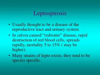
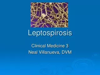
Background • Caused by gram negative spirochetes: Genus Leptospira • Most common zoonosis (disease from animals) in the world • Humans and animals (mammals, birds, amphibians and reptiles) can develop Leptospira infection • Humans are rarely chronic carriers (they are accidental hosts) • Animals are often chronic carriers
A scanning electron micrograph depicting Leptospira on a 0.1-µm polycarbonate filter
How does infection happen? • Direct contact with • the body fluid of an acutely infected animal • exposure to soil or fresh water contaminated with the urine of an animal that is infected or a chronic carrier
How does infection happen? • Leptospires enter host through… • abrasions in healthy skin • waterlogged , wet skin • intact mucus membranes or conjunctiva, nasal mucosa • lungs (aerosolized body fluid) • placenta (during pregnancy) • Then through the lymphatics, causing septicemia and spread to all organs • Host develops immunoglobulin to clear bacteria
Two stages • Acute • septicemic stage of acute febrile illness • lasts 5-7 days • Delayed • immune phase • lasts 4-30 days
Two presentations • Anicteric leptospirosis • self limited disease, flu like illness • septicemic phase (acute) • immune phase (delayed) • Icteric leptospirosis (Weil disease) • severe illness with multi-organ involvement and failure • septicemic and immune phase are mixed together
Morbidity/mortality • Mild forms are rarely fatal • 90% of cases are mild • In severe forms, mortality is 5-40% • Elderly and immunocompromised are at highest risk for severe cases • Working aged-males have more exposure
How quickly and for how long? • Incubation period usually 5-14 days (3-30) • Leptospires remain in renal tubules, brain anterior chamber of eye for 60 days (longer in animals)
Leptospirosis is an infective systemic vasculitis • Leptospires multiply in the small blood vessel endothelium resulting in vasculitis
Leptospirosis is an infective systemic vasculitis • Kidneys • interstitial nephritis and tubular necrosis • Liver • centrilobular necrosis and hepatocyte dysfunction • Pulmonary • alveoloar and interstitial vascular damage with hemorrhage ( MAJOR CAUSE OF LEPTOSPIROSIS ASSOCIATED DEATH )
Leptospirosis is an infective systemic vasculitis • Skin • epithelial blood vessels leak (rash, purpura) • Skeletal muscle • edema • Capillary leakage • hypovolemia, shock • Coagulation abnormalities • disseminated intravascular coagulation (DIC) • hemolytic uremic syndrome (HUS) • thrombotic thrombocytopenic purpura (TTP)
Up to 80% of people in the tropics have evidence of past infection
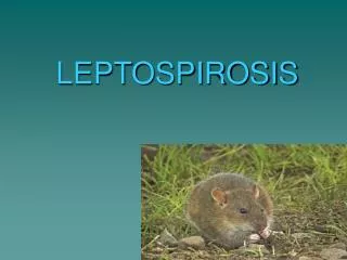
History • Exposure • Urine, kidneys, or conception products of infected animals. • Rodents, dogs, cattle and pigs • Direct infection (body fluids, organs of infected animals) • Indirect infection (inoculated soil and water)
Acute phase 5-7 days • High temperature • Rigors • Sudden headache • Nausea and vomiting • Anorexia • Diarrhea • Cough, pharyngitis • Non pruritic skin rash • Muscle pains (calf and lumbar area)
Physical • Fever (about 7 days) • Signs of volume depletion • tachycardia • hypotension • oliguria • Abnormal chest exam • Skin • transient petechial eruption (palate) • Jaundice • purpura • Eyes • conjuctival suffusion (like conjunctivitis but no exudates) • uveitis
Physical • Muscle tenderness • paraspinal and calf muscles • Lung exam • crackles or consolidation from alveoloar hemorrhage • Myocarditis • signs of heart failure • Abdomen • liver enlargement and tenderness from acalculouscholecystitis • Meningeal signs • neck stiffness • rigidity • photophobia • delerium • In severe disease, may have prolonged mental symptoms like depression, anxiety, irritability, psychosis, dementia
Clinical Diagnosis Fever > 38.0°C (100.4 °F) AND at least TWO from COLUMN A or ONE from COLUMN B • COLUMN A • Headache • Myalgias • Jaundice • Chills/Rigors • Skin rash • Conjunctival suffusion without purulent discharge • COLUMN B • Aseptic meningitis • Acute renal failure • Hemorrhagic pneumonitis • Cardiac arrhythmias, EKG abnormalities • Liver failure • Jaundice with acute renal failure (Weil’s disease)
Differential diagnosis Influenza Enteric fever Hantavirus Rickettsial disease Encephalitis Primary HIV infection
Laboratory: confirm diagnosis • Isolate the leptospires from human tissue or body fluids (urine) • Paired acute and convalescent serum specimens using microscopic agglutination test (MAT) 4X rise in antibody titre • Singly MAT titer of 1:800 • Identification of spirochetes on dark-field microscopy
Laboratory: organ involvement • CBC (hemorrhage, platelets from DIC) • Creatinine and BUN (renal failure, shock) • Magnesium and potassium (wasting) • Bilirubin, (capillaritis in the liver). Alkaline phosphatase AST, ALT • INR PTT (DIC, liver disease)
Diagnostic imaging • Chest Xray • bilateral diffuse airspace disease • cardiomegaly, pulmonary edema • patchy infiltrates • Abdominal ultrasound • acalculous cholecystitis
Treatment: antimicrobial • Outpatient treatment • Oral doxycycline • Decreases duration of fever and most symptoms • 100 mg PO bid for 7 days • Hospitalized patient • Intravenous penicillin G • 20-24 million U/d IV divided q4-6h for 7 days • Intravenous doxycycline • 100 mg IV q12h for 7 days
Other antimicrobials • Amoxicillin • 0.5-1.0 g PO q8h • Cefotaxime • 1 g IV q6h • Ceftriaxone • 1 g IV q24h • Erythromycin (pregnant, penicillin allergy) • 500 mg IV q6h • 500 mg PO qid
Treatment: supportive • Monitor and treat patient for • Shock • Renal failure (usually always reversible) • Respiratory failure (mechanical ventilation) • Cardiac monitoring (arrhythmias) • Plasma exchange, corticosteroids, IV immunoglobulin
Prevention • Avoid or reduce contact with animals, contaminated soil or water • Wear protective garments including footwear, gloves and eye protection • Attention to hygiene and sanitation
What new knowledge do you have? • Write down different information! • 2 animals that can be infected with leptospirosis • 2 ways the bacteria can enter the human body • 2 complaints the patient may have (history) • 2 examination findings (physical) • 2 organs that may be involved and how the organ damage can be shown • 2 antibiotics to treat leptospirosis