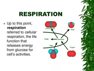
RESPIRATION
230 likes | 251 Vues
Learn about the transport of oxygen (O2) and carbon dioxide (CO2) in the blood through hemoglobin. Understand the O2-Hb dissociation curve and how it affects loading and unloading reactions. Explore factors that influence the affinity between Hb and O2.

RESPIRATION
E N D
Presentation Transcript
RESPIRATION Dr. Zainab H.H Dept. of Physiology Lec. 8
objectives • Discriminate between O2 & CO2 transport. • Describe O2- Hb dissociation curve
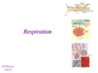
Hb and O2 Transport • Each Hb has 4 polypeptide (2 α and 2 b) chains, each combined with heme group. • In the center of each heme group is 1 atom of iron that can combine with 1 molecule O2. • One Hb molecule thus combine with four O2molecules. • There are about 280 million Hb molecules/RBC • each RBC can carry over a billion molecules of O2
Hemoglobin • Oxyhemoglobin: • Normal heme contains iron in the reduced ferrous form (Fe2+). • Fe2+ shares electrons and bonds with O2. • Deoxyhemoglobin: • When oxyhemoglobin dissociates to release O2 to the tissues, heme iron is still in the reduced form. • Hb does not lose an electron when it combines with O2.
Hemoglobin (continued) • Methemoglobin: • Has iron in the oxidized ferric form (Fe3+). • lacks the electron it needs to form a bond with O2 and cannot participate in O2 transport. • Blood normally contains a small amount. • Carboxyhemoglobin: • The reduced heme is combined with CO. • The bond with CO is 210 timesstronger than the bond with O2. • Transport of O2 to tissues is impaired.
Hb Concentration • O2-carrying capacity of blood determined by its Hb concentration: • In anemia: • Hb concentration below normal abnormally low O2content of the blood. • In polycythemia: • Hbconcentration above normal abnormally low O2content of the blood.
Hb Concentration • Erythropoietin: hormone produced by the kidneys controls production of RBCs in the bone marrow. • If the amount of O2 delivered to the kidneys is lower than normal stimulates secretion of erythropoietin production of RBCs. • Androgens: promotes RBCs production • explains why the Hb concentration in men is from 1 to 2 g per 100 ml higher than in women.
The Loading and Unloading Reactions • Deoxyhemoglobin + O2 Oxyhemoglobin (loading reaction) occurs in the lungs • Oxyhemoglobin deoxyhemoglobin + O2 (Unloading reaction) occurs in the tissues. • Loading and unloading shown as a reversible reaction: (tissues) Deoxyhemoglobin + O2 Oxyhemoglobin (lungs)
The Loading and Unloading Reactions • The extent to which the reaction will go in each direction depends on: • PO2 of the environment • High PO2 drives the equation to the Rt. (favors the loading reaction). • Low PO2 in the systemic capillaries drives the reaction in the opposite direction (promote unloading). • The extent of this unloading depends on how low the PO2 values are.
The Loading and Unloading Reactions • affinity (bond strength) between Hb and O2. • Very strong bond would favor loading but inhibit unloading • Weak bond would hinder loading but improve unloading. • The bond strength between Hb and O2 is normally strong enough so that 97% of the Hb leaving the lungs is in the form of oxyHb. • BUTthe bond is sufficiently weak so that adequate amounts of O2 are unloaded to sustain aerobic respiration in the tissues.
O2 - Hb Dissociation Curve (OHDC) • At PO2 of 100 mmHg, blood in the systemic arteries, has a percent oxyHb saturation of 97% (97% of the Hb is in the form of oxyHb) delivered to the systemic capillaries O2 diffuses into the cells and is consumed in aerobic respiration. • Blood leaving the systemic veins is reduced in O2; it has a PO2 of about 40 mmHg and a percent oxyHb saturation of about 75%.
OHDC (continued) • Expressed another way: • gram of Hb carry about 1.34ml of O2, so if Hb were 100% saturated, it carry about 20ml of O2 • normally it is only about 97-98% saturated so it actually carries about 19.4ml of O2 • Blood entering the tissues contains 20 ml O2/100 ml blood, and blood leaving the tissues contains 15.5 ml O2/100 ml blood (14.4). • Thus, 22%, or 4.5 ml of O2out of the 20 ml of O2 per 100 ml blood, is unloaded to the tissues.
OHDC (continued) • The relatively large amount of oxyHb remaining in the venous blood at rest serves as an O2 reserve. • If a person stops breathing, a sufficient reserve of O2 in the blood will keep the brain and heart alive for about 4 to 5 minutes without using cardiopulmonary resuscitation (CPR) techniques. • This reserve supply of O2 can also be tapped when a tissue’s requirements for O2 are raised.
OHDC (continued) • A graphic illustration of the percent oxyH saturation at different values of PO2 • The values in this graph are obtained by subjecting samples of blood in vitro to different PO2. • The percent oxyHb saturations obtained can be used to predict what the unloading percentages would be in vivo with a given difference in arterial and venous PO2 values.
OHDC (continued) • It is S-shaped, or sigmoidal due to Heme- Heme interaction. • Plateau portion of the curve: • Range that exists at the pulmonary capillaries. • relatively flat at high PO2 values indicates that changes in PO2 within this range have little effect on the unloading reaction. • Minimal O2 transported until the PO2 falls below 60 mm Hg.
OHDC (continued) • Steep portion of the curve: • Range that exists at the systemic capillaries • small changes in PO2 produce large differences in % saturation (unload more O2)
A 36-year-old woman is found comatose at her home and is life-flighted to the nearest regional medical center. Blood gases reveal a normal PaO2 but a lower-than normal arterial O2 saturation. Which of the following conditions is most consistent with the findings? • a. Anemia • b. Carbon monoxide poisoning • c. Hypoventilation • d. Low V/Q ratio • e. Right-to-left shunt