Carbohydrates Chapter 20
Carbohydrates Chapter 20. Fig. 17-CO, p.531. Have basic formula ratio of C 1 H 2 O 1 1C to 1H 2 O Carbo Hydrate. Carbohydrates Carbohydrates are polyhydroxy aldehydes or ketones, or substances that yield such compounds upon hydrolysis. Glucide is the French word for carbohydrate

Carbohydrates Chapter 20
E N D
Presentation Transcript
Have basic formula ratio of C1H2O1 1C to 1H2O Carbo Hydrate Carbohydrates Carbohydrates are polyhydroxy aldehydes or ketones, or substances that yield such compounds upon hydrolysis. Glucide is the French word for carbohydrate Example:
Classification of Carbohydrates Carbohydrates are classified according to size: Monosaccharide – a single polyhydroxy aldehyde or ketone unit. We can also call these the base sugar units. Disaccharide – composed of two monosaccharide units Oligosaccharides – 3-10 monosaccharide units Polysaccharides – very long chains of linked monosaccharide units. Typically hundreds, thousands or even millions of units long Glycosides-sugars attached to other groups, the sugar part is called a glycone (one to many units).
Classified as to being digestible • This refers to being digestible by humans (either themselves or by gut bacteria) • Glucide- digestible (japan) • Glucide- • Simple carbohydrates (sugars mono and di) • Complex carbohydrates (starches) • Fiber (not digestible) typically cellulose • Other (not digestible)
Stereochemistry Many carbohydrates exist as enantiomers – or stereoisomers that are mirror images.
A chiral object cannot be superimposed on its mirror image. A chiral carbon is one that has four different groups attached to it.
The presence of a single chiral carbon gives rise to stereoisomerism. If a carbon atom is attached to four different groups, it is chiral. If any two groups are identical, it is not chiral.
Compounds can have more than one chiral carbon:• The maximum number of stereoisomers is 2n where n= number of chiral carbon atoms.
Fischer Projections Fischer projections depict three-dimensional shapes for chiral molecules, with the chiral carbon represented by the intersection of two lines.
D will have OH to right Fisher projections of carbohydrates have the carbonyl (C=O) at the top. The hydroxyl group on the chiral carbon farthest from the C=O group determines whether the carbohydrate is D (OH on right) or L (OH on left). Flag
In the Fischer projection the last OH is to the right in “D” D and L Enantiomers D = Dextro – Right handed sugar – from bottom of Fisher projection the second OH up is on the right L = Levo – Left handed sugar – from the bottom of Fisher projection the second OH up is on the left L D
D and L enantiomers rotate polarized light in opposite directions.
The enantiomer that rotates polarized light to the left is the levorotatory or (-) enantiomer. The enantiomer that rotates it to the right is the dextrorotatory or (+) enantiomer. The D and L designations do not represent dextrorotatory and levorotatory. Each chiral group can rotate the light, so the net rotation is the combination of rotations from each. D and L only tell how the bottom chiral group rotates the light. So is possible for a D sugar to have a (-) (net rotation to the left)
In some instances only the D or L enantiomers are found in nature. They are rarely found together in the same biological system. Almost all biologic sugars are only the D-isomer. (amino acids are virtually only in the L form for all organisms) • For example, humans can only metabolize the D-isomers of monosaccharides. • Only the D-isomer sugars are sweet
Monosaccharide Classification Classified by whether the monosaccharide is an aldehyde (aldose) or ketone (ketose). Classified by the number of carbon atoms in the monosaccharide.
Carbon at crossed lines Combining Monosaccharide Classifications These are Fischer Projections ( the carbons are rolled away from you and the side groups are toward you)
Fisher projections Fig. 17-8, p.542
Physical Properties of Monosaccharides Because of the many –OH groups, they form hydrogen bonds with water molecules and are extremely water soluble. Most are called sugars because they taste sweet.
Physical Properties Monosaccharides are colorless crystalline solids, very soluble in water, but only slightly soluble in ethanol Sweetness relative to sucrose:
Monosaccharide Reactions All monosaccharides with at least five carbon atoms exist predominantly as cyclic hemiacetals and hemiketals. A Haworth structure is a standard way of writing the sugars in the cyclic (ring) form A Haworth structure can be used to depict the and anomers of a monosaccharide. Anomers are stereoisomers that differ in the 3-D arrangement of groups at the anomeric carbon of an acetal, ketal, hemiacetal, or hemiketal group.
1 1 FischerProjection(linear form) 1 1 -(beta)OH up HayworthProjection(ring form) -(alpha)OH down
Fisher Diagram Haworth Diagram Haworth Diagram Likes this form because it can H-bond to the Flag OH p.544a
Monosaccharide Reactions, cont. A reducing sugar can be easily oxidized. All aldose monosaccharides are reducing sugars. Benedict’s reagent tests for the presence of reducing sugars: Reducing sugar + Cu2+ oxidized compound + Cu2O blue orange-red precipitate IGNORE - skip
Ester linkage Notice the phosphate is connected to the carbon # 6 Monosaccharide Reactions, cont. The –OH groups of monosaccharides can behave as alcohols and react with acids (especially phosphoric acid) to form esters.
Monosaccharide Reactions, cont. Cyclic monosaccharide hemiacetals and hemiketals react with alcohols to form acetals and ketals, referred to as glycosides.
We don’t need for chapter 22- we will cover in Ch 26 Important Monosaccharides 5 Carbon Sugars Ribose and Deoxyribose Used in the synthesis of DNA and RNA. 6 Carbon Sugars Glucose Most nutritionally important monosaccharide Sometimes called dextrose or blood sugar Galactose A component of lactose (milk sugar) Fructose The sweetest monosaccharide Sometimes called levulose or fruit sugar A ketose
Disaccharides Two monosaccharide units linked together by acetal or ketal glycosidic linkages.
a(1 4) glycosidic linkage O Disaccharides Two monosaccharide units linked together by acetal or ketal glycosidic linkages. b(1 4) glycosidic linkage O
Important Disaccharides Maltose Two glucose units linked (14) Formed during the digestion of starch to glucose Lactose Galactose and glucose units linked (14) Found in milk Sucrose Fructose and glucose units (1 b2) Found in many plants (especially sugar cane, sugar beets) Not a reducing sugar
Glycosides • One to many sugar groups attached to other molecules (the sugar part is called the glycone and the non sugar part the genin) • Glycoproteins: many times membrane bound and may be used for cell recognition and response (ex blood types • Glycolipids: typically membrane bound and may be used for flags to signal cellular need • Small molecules bound to sugar units may inactivate molecules for storage or make them soluble for elimination
Glycocalyx(a glycoprotein) • A glycocalyx, literally "sugar coat", is a network of polysaccharides that project from cellular surfaces, e.g. those of bacteria. It serves to protect the bacterium by creating capsules, or allows the bacterium to attach itself to inert surfaces (like teeth or rocks; e.g. Streptococcus pneumoniae attaches itself to lung cells), eukaryotes, or other bacteria (their glycocalyxes can fuse to envelop the colony). • Its presence on inert materials (such as metal hardware implanted for fracture fixation or total joint replacement) make it difficult to eradicate deep infections as the bacteria will 'cling' on to the material via the glycocalyx. It is therefore often necessary to completely remove the hardware device in order to fully eradicate a wound infection.
Human Glycocalyx • The glycocalyx is also the name given to a specific structure of a mature platelet. The glycocalyx is unique among the cellular components of the blood. It is similar to the bacterial glycocalyx above in that it is made up of glycoproteins and allows the platelet to adhere to surfaces such as collagen of damaged vessels. The glycocalyx appears as a fluffy coat to the outer membrane of platelets and contains many of the receptor proteins that allow cell adhesion. Glycocalyx also appears on the cells lining blood vessels (endothelial cells). Among its established roles are reducing friction to the flow of blood and serving as a barrier for loss of fluid through the vessel wall. In times of inflammation, the endothelial cell glycocalyx is sheared off, to permit attachment of leukocytes and movement of water from microvessels. • The glycocalyx is chemically unique in everyone (cellular fingerprints) but identical in monozygote twins, and acts like an identification tag that enables the body to distinguish its own healthy cells from transplanted tissues, invading organisms and diseased cells. Human blood types and transfusion compatibility are determined by glycolipids and glycoproteins. • A glycocalyx can also be found on the apical portion of microvilli within the digestive tract, especially within the small intestine. It creates a meshwork 0.3 micrometers thick and consists of acidic mucopolysaccharides and glycoproteins that project from the of epithelial absorptive cells. It provides additional surface for adsorption and includes enzymes secreted by the absorptive cells that are essential for the final steps of digestion of proteins and sugars.
Human Glycocalyx Functions • Protection: Cushions the plasma membrane and protects it from chemical injury • Immunity to infection: Enables the immune system to recognize and selectively attack foreign organisms • Defense against cancer: Changes in the glycocalyx of cancerous cells enable the immune system to recognize and destroy them • Transplant compatibility: Forms the basis for compatibility of blood transfusions, tissue grafts, and organ transplants • Cell adhesion: Binds cells together so that tissues do not fall apart • Inflammation regulation: Glycocalyx coating on endothelial walls in blood vessels prevents leukocytes from rolling/binding in healthy states (sheared off in inflamed tissue cells) • Fertilization: Enables sperm to recognize and bind to eggs • Embryonic development: Guides embryonic cells to their destinations in the body
Polysaccharides(from a dietary perspective) • Digestible-complex carbohydrates- starch-storage carbohydrates • 1. grains-culturally different • wheat- European • Rice- East Asian • Corn- Latin America • 2. Bean and Pea Family • 3. Tubers (potatoes, yams, cassava) • 4. Very little from leaves or meat • Indigestible-Fiber or ruffage • (Note: complex carbohydrates is not a scientific but a nutritional term. Usually meant to be digestible but sometimes is applied to all polysaccharides in our diet
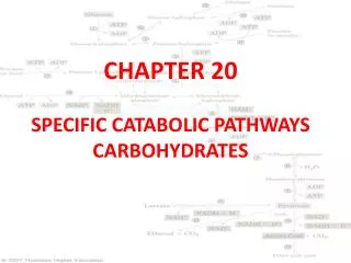
Polysaccharides StarchComplexes with iodine to form a dark blue color A polymer consisting of glucose units Has two forms in plants and 1 form in animals PLANTS Amylose: Unbranched (plants) Amylopectin: Branched (plants) (every 24-30 units) ANIMALS Glycogen: Branched (animals) (every 8-12 units), more highly branched Each connection is a glycosidic bond
The Structure of Amylose Linear, no branches
Polysaccharides, cont. Glycogen (animal starch) – a polymer of glucose units.Used to store glucose, especially in the liver and muscles.Structurally similar to amylopectin with (14) mainly and (16) linkages at branch points, but glycogen is more highly branched (about every 8-12 units is a branch)
A glycogen storage site on the surface of the Phosphorylase enzyme binds the glycogen particle. Given the distance between storage & active sites, Phosphorylase can cleave a(14) linkages only to within 4 residues of an a(16) branch point. This is called a "limit branch". Explore the structure of muscle Glycogen Phosphorylase with Chime.
Debranching enzyme has 2 independent active sites, consisting of residues in different segments of a single polypeptide chain: • The transferase of the debranching enzyme transfers 3 glucose residues from a 4-residue limit branch to the end of another branch, diminishing the limit branch to a single glucose residue. • The a(16) glucosidase moiety of the debranching enzyme then catalyzes hydrolysis of the a(16) linkage, yielding free glucose. This is a minor fraction of glucose released from glycogen. • View an animationhttp://www.rpi.edu/dept/bcbp/molbiochem/MBWeb/mb1/part2/glycogen.htm The major product of glycogen breakdown isglucose-1-phosphate, from Phosphorylase activity.