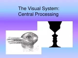
Visual Processing
Visual Processing. Sensory Neural Systems 5 February 2008 Rachel L. Le ón. Review. Basic eye structure Sensory cells of the eye Retinal processing. http://scienceblogs.com/clock/2006/06/bio101_lecture_6_physiology_re.php.

Visual Processing
E N D
Presentation Transcript
Visual Processing Sensory Neural Systems 5 February 2008 Rachel L. León
Review • Basic eye structure • Sensory cells of the eye • Retinal processing http://scienceblogs.com/clock/2006/06/bio101_lecture_6_physiology_re.php http://www.chemistry.wustl.edu/~edudev/LabTutorials/Vision/images/Rodcell.jpg http://www.matossianeye.com/art2/ANATONY2.jpg
Neural Coding in the Retina • Ganglion cells influenced by many receptors • Pattern of illumination that maximally excites ganglion cell is doughnut shaped • Center-surround receptive field • Lateral inhibition of receptive fields enhances boundaries • X, Y, W ganglion cells http://www.neurobio.arizona.edu/282/Lectures2005/Visual%20pathways/Blk3Lec2.htm
Outline • Classical Visual Pathway • Organization of optic nerves and tracts • Lateral geniculate nuclei • Primary visual cortex • Other Pathways http://www.nature.com/nrn/journal/v6/n3/fig_tab/nrn1630_F4.html
Optic Nerve and Tract • Temporal retinal fibers maintain ipsilateral position • Nasal retinal fibers cross at optic chiasm • Optic tract contains all information from contralateral visual field http://library.thinkquest.org/26313/eye_wo1.jpg
Lateral Geniculate Nucleus • LGN receives most input from optic tracts • 6 layer structure • Magnocellular layers • Parvocellular layers http://www.physics.utoledo.edu/~lsa/_color/17_eye.htm
Gross Dissection – Human Visual Pathway http://anatomy.uams.edu/AnatomyHTML/atlas_html/eye_38.html
Primary Visual Cortex • Simple cells vs. Complex cells • Cortical orientation columns • Ocular dominance columns • Blobs – receive parvocellular input http://www.neurobio.arizona.edu/282/Lectures2005/Visual%20pathways/10_022.jpg
Other Visual Pathways • Most of the axons not going to LGN terminate in superior colliculus • Some efferents from SC go to pulvinar • Pulvinar efferents terminate in visual cortex other than V1 • Some optic tract axons terminate in pretectal area