Wrist & Hand
Wrist & Hand. Long Nguyen. Past exam q’s.

Wrist & Hand
E N D
Presentation Transcript
Wrist & Hand Long Nguyen
Past exam q’s • Diagram of a cross section of Radius, Ulna and tubes provided with the caption: “At the lower end of the forearm there is an extensor retinaculum. Septa attach the retinaculum to the radius and ulna forming six osseofascial tunnels for the extensor tendons”. List the tendons corresponding to each of the numbered tunnels above. (Oct 00) • What is the arrangement of structures passing from the forearm to the wrist at the level of the carpal tunnel within the flexor retinaculum. • Write short notes on the anatomy of the lunate bone. (Sept 04) • Draw a clear diagram and anatomical relations of the scaphoid bone. (April 04) • Write notes on the supernumerary bones of the hand. (?) • In the embryological development of the hand and foot, many of the bones are homologous (for example the radius is a homologue of the tibia and the ulna is homologous to the fibula). Which carpal bones and tarsal bones are homologues? (Sept 02) • Regarding the thumb: (Oct 01) a what muscles are responsible for thumb flexion? b extension? c abduction? d adduction? e what is the nerve supply of each of these muscles • Write short notes on the arterial supply to the hand. (April 04)
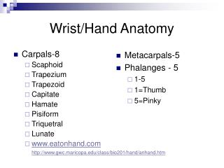
Bones of the hand • 8 carpal bones, 5 metacarpal bones, 14 phalanges • 8 carpal bones lie in two semicircular rows separated by S-shaped midcarpal joint • Prox row – S, L, Tqm, P • Distal row – Tpzm, Tpzd, C, H • Carpal bones articulate with each other by intercarpal joints
Carpal bones • Scaphoid bone (boat shaped) • Most commonly fractured carpal bone • Convex articular proximal surface for radius • Flat surface medially for lunate • Concave distomedially for capitate • Convex distal articulation with trapezium & trapezoid • Tubercle on the volar aspect of distal lateral surface • Narrow waist perforated by vascular foramina more numerous distally • In 15%, blood supply to scaphoid is solely supplied from nutrient arteries which pass distal to proximal • Fracture trhough the waist can produce avascular necrosis of proximal portion • Lunate • Most commonly dislocated carpal bone (displaced anteriorly) • Convex proximal facet for radius • Concave distally for capitate • Facet for adjoining bones on either side
Carpal bones • Triquetral • Oval fact on distal palmar surface for pisiform • Pisiform • Flat surface for articulation with triquetral • Trapezium • Saddle-shaped distal articular surface with 1st MC • Tendon of flexor carpi ulnaris lies in a vertical groove on the palmar surface • Trapezoid • Capitate • Convex proximal surface for lunate articulation • Articulates with 3rd MC and small facet for 4th MC • Hamate • hook projects from the distal part of the palmar surface, and is directed laterally
Metacarpals & phalanges • 1st MC (saddle joint) articulates solely with trapezium • Remaining MC’s had expanded bases and articulate with each other and the distal row of carpal bones • Middle MC has prominent styloid process projecting dorsally into angle between trapezoid and capititate • MCJ and IPJ are hinged synovial joints
Ossification • Carpus cartilagenous at birth • Ossification in a clockwise direction from capitate • Capitate & hamate 1 year • Triquetral 2-3 year • Lunate 3-5 year • Scaphoid, trapezium, trapezoid: 6 year • Pisiform 10-12 year • MC & phalangeal shafts ossify in utero • Radiographs of the left hand are obtained for bone age assessment by comparing features such as epiphyseal appearance and fusion
18m 3y 9m 5y 4m 7y 1m 13y
Ossification - variable timing 2y 11m 5y 4m 6y 11m 7y 1m
Supernumery bones • Sesamoid in the tendon of the flexor pollicis brevis near MC head of thumb • Os centrale found between scaphoid, trapezoid and capitate. May represent tubercle of scaphoid that has not fused with upper pole • Os radiale externum immediately distal to radial styloid
Wrist Joint • Condyloid/ellipsoid synovial joint • Radius articulates with scaphoid and lunate. Articulation with triquetral only on ulnar deviation • Ulna shorter than distal radius. Fibrocartilaginous disc projects laterally from ulna styloid to the radius. It articulates with lunate and triquetral • Fibrous capsule lined by synovium incloses the joint • Strengthened by dorsal and palmar radiocarpal ligaments which run distally and medially from radius • Radial and ulnar collateral ligaments
Wrist Movements • Flexion (80º) • Flexor carpi radialis & flexor carpi ulnaris • Palmaris longus, flexors of fingers and thumb, abductor pollicis longus • Extension (60º) • Extensor carpi radialis longus, extensor carpi radialus brevis, extensor carpi ulnaris • Extensors of fingers and thumb • Abduction (15º radial) • FCR, ECRL, ECRB • APL • Adduction (45º ulna) • FCU, ECU
Extensor retinaculum • Antebrachial fascia thickened posteriorly at the wrist to form a transverse 2.5cm band • Proximal attachment: anterolateral border of radius above styloid process • Distal attachment: pisiform & triquetral • 6 shealths containing 9 tendons occupy the six osseofibrous tunnels deep to the extensor retinaculum • A) 3 for thumb in 2 shealths (EPL) (EPB,APL) • B) 3 for extensors of the wrist in 2 sheaths (ECU) (ECRL,ECRB) • C) 3 for extensors of the digits in 2 sheaths (EDigitorum, E Indicis) (Extensor digiti minimi)
Palmar Aponeurosis • Fascia of the forearm continues distally as the deep fascia of the palm • Fascia is thickened in the palm (thin over the thenar and hypothenar eminences) • Overlies long flexor tendons of the palm • Proximal end continuous with flexor retinaculum • Distal ends of the aponeurosis divides at the roots of the digits into four longitudinal bands which attaches to base of prox phalanx
Fascial compartments • Central compartment • Flexor tendons and shealths • Superficial palmar arch • Branches of median and ulnar nerves • Superior boundary: palmar aponurosis • Inferior boundary: deep muscles of palm (adductor pollicis) • Bounded on either side by septa passing from medial and lateral edges of aponeurosis to 1st and 5th MC bones • Medial/hypothenar compartment • Lateral thenar compartment
Potential space between central compartment and the deep muscles of the palm: midpalmar space • Retroadductor space between adductor pollicis and 1st dorsal interosseous muscle
Carpal tunnel • Carpal bones angled producing anterior concavity • Hollow formed by these bones is bounded anteriorly by flexor retinaculum • Flexor retinaculum attachments • Medially: hook of hamate & pisiform • Laterally: ridge of trapezium & scaphoid tubercle • Contents: • Median nerve immediately deep to retinaculum • Flexor pollicis longus tendon • Tendons of flexor digitorum superficialis (x4) and flexor digitorum profundus (x4) • Tendon of flexor carpi radialis grooves the trapezium and lies in separate compartment of tunnel • Relations • Ulnar artery superficial to flexor retinaculum, lateral to ulnar nerve
Intrinsic hand muscles • Palmar aspect • 3 groups • Thenar muscles • Hypothenar muscles • Lumbricals and interossei
Thenar muscles • Produce thenar eminence • Oppose thumb • Supplied by recurrent branch of median nerve
Short muscles in the hand • 4 Lumbricals • act on medial 4 digits • Flex digits at the MCPJ and extend IPJ • 7 Interossei • 3 palmar adduct (PAD) • 4 dorsal abduct (DAB)
Long flexor tendons • Flexor digitorum superficialis tendon • Enters the fibrous flexor shealth on the palmar surface of the flexor digitorum profundus • Splits into 2 and wraps around the profundus and meets on deep surface in a chiasma • Distal to chiasma, superficialis tendon is attached to margins of the front of middle phalanx • Profundus tendon • enters fibrous sheath deep to the superficialis • then lies superficial distal to the split of the superficialis tendon • attaches to base of terminal phalanx • Each tendon receives blood vessels invested in synovial membrane called vincula
Long extensor tendons • Extensor tendon blends in with a triangular fibrous expansion on the dorsum of the proximal phalanx • Margins of expansion thickened by attachments of the tendons of lumbrical and interossei • Extensor tendon splits into middle and two collateral slips as it approaches PIPJ • Middle slip attached to base of middle phalanx • Collateral slips joined by thickened margins of expansion and converge to insert into base of distal phalanx
Arterial supply • Ulnar artery continues as superficial palmar “arch” • Usually not complete arch • Continuous with superficial palmar branch of radial artery if complete • Gives rise to 3 common palmar digital arteries which run distally to the webs between the fingers and divides into proper palmar digital arteries (run alongside 2nd-4th digits) • Radial artery • Princeps pollicis artery (thumb) • Radialis indicis artery (lateral side of 2nd digit) • Joins deep branch of ulnar artery to form deep palmar arterial arch • Arch gives rise to 3 palmar metacarpal arteries which run distally and join the common palmar digital arteries