Nephrology 2
Nephrology 2. Euan Green Mr Betts. Assessment of renal function. History and examination clues Blood tests and associated formulae Urine tests incl. 24h clearance Nuclear medicine techniques Chronic renal failure 1)How does autoregulation of renal blood flow work?

Nephrology 2
E N D
Presentation Transcript
Nephrology 2 Euan Green Mr Betts
Assessment of renal function • History and examination clues • Blood tests and associated formulae • Urine tests incl. 24h clearance • Nuclear medicine techniques • Chronic renal failure • 1)How does autoregulation of renal blood flow work? • 2) Why do you get hypertension in chronic renal failure? • 3) What are the implications of chronic renal failure?
MCQ • CKD stage 5 equates to an eGFR <5mls/min • CKD stage 1 equates to an eGFR 75-90mls/min • Those identified with CKD stage 2 should be referred to a nephrologist • The CKD classification is based solely on eGFR • CKD stage 3 has been subdivided into those with an EGFR < or >= 45mls/min
MCQ • CKD stage 5 equates to an eGFR <5mls/min • CKD stage 1 equates to an eGFR 75-90mls/min • Those identified with CKD stage 2 should be referred to a nephrologist • The CKD classification is based solely on eGFR • CKD stage 3 has been subdivided into those with an EGFR < or >= 45mls/min
Chronic kidney disease Kidney damage= persistent microalbuminuria, persistent proteinuria, persistent haematuria, structural abnormalities of the kidneys demonstrated on ultrasound scanning or other radiological tests, or biopsy-proven chronic glomerulonephritis)
Why should we detect kidney disease • The scale of the problem • 0.1% of the population on dialysis (40,000 in the UK) • Costing the NHS an average of £20,000 per patient per year (3% of the NHS budget) • 4.5% of the population have moderate or severe renal failure but not yet on dialysis • Patients with renal failure are considered to be in the highest risk category for heart disease (20% per 10 years)
Death • Patients with CKD are more likely to diethan require dialysis • 27,998 CKD patients followed for 5 years: Keith DS, AIM 2004;164:659-663
Poor outcome of unreferred CKD • East Kent study 601,000 population • Using opportunistic serum creatinine • Males - serum creatinine 180 mol/L • Females - serum creatinine 135 mol/l • Approximate to GFR < 30-40ml/min/1.73m2 • Prevalence 4708 unreferred • Outcome (over 31 months) • Median survival 28 months • Cardiovascular 40% • Infection 26% • Cancer 16% • End stage renal failure <5%
Case 1 • A 45 year old man has been referred for a vasectomy under GA • His pre-op bloods show renal impairment (eGFR 70) and he’s brought back to clinic to assess this unexpected finding. • How would you assess for evidence of renal dyfunction?
Case 1 • History • Age • Co-morbidities • Hypertension • Diabetes • Vascular disease • Obesity • Smoking • Structural urinary tract abnormalities • Liver disease • Cancers
Case 1 • Concurrent illness • Dehydration from D&V • Recent URTI • Recent surgery • Drugs • Lots • Remember X-ray contrast and other once offs • Family history
Case 1 • Examination • Blood pressure • Abdominal masses (bladder, kidneys, ascites) • Signs of fluid retention (peripheral oedema, pulmonary oedema, JVP) • Fluid balance • Changes in weight
Bicohemistry Tests • Tests • Urine dipstick • Urine tests • Microscopy • 24 hour urine collection • Urinary Albumin:creatinine ratio • Blood tests
MCQ • All proteins except albumin cause a colour change on urine dipstick • Prolonged periods of standing can underestimate proteinuria • A urinary albumin:creatinine ratio of 25mg/mmol is normal • Creatinine clearance is always an overestimate of GFR • Creatinine is freely filtered at the glomerulus and neither secreted, nor reabsorbed
MCQ • All proteins except albumin cause a colour change on urine dipstick • Prolonged periods of standing can underestimate proteinuria • A urinary albumin:creatinine ratio of 25mg/mmol is normal • Creatinine clearance is always an overestimate of GFR • Creatinine is freely filtered at the glomerulus and neither secreted, nor reabsorbed
Case 2 • Friday at 20:05 On call referral: • A man has pitched up in A&E with a note. • “Dear urologist, This 60 year old chap had dipstick haematuria so I sent him for a CT urogram yesterday. The report says ‘mild bilateral hydronephrosis, images in the pelvis are uninterpretable due to streak artefact from bilateral hip replacements. Clinical correlation advised’. His creatinine is 150 today. Please clinically correlate as discussed with your SHO.”
Case 2 • A&E have helpfully done a urine dipstick which shows • Blood ++ • Protein ++ • They’ve done a post void bladder scan which is normal • They’ve confirmed the creatinine result • In doing so he’s reached 3 hours and 59 minutes in the department and the bed manager has admitted him under urology after discussing with the SHO.
Urine dipstick • Protein • Tetrabromophenol reaction with albumin causes a colour change (yellow to blue/green) • Trace 5-20mg/dL • + ~30mg/dL • ++ ~100 mg/dL • +++ ~300mg/dL • ++++ >2000mg/dL • False +ve • Orthostatic proteinuria • Iodinated contrast • Alkaline urine • Blood • Peroxidase reaction • (See BAUS consenus statement on haematuria)
Case 2 • You take a history and examine him and there’s nothing to find • He’s a medical negligence lawyer whose best friends is your trusts chief exec. and refuses to go home until this is all sorted. • The med reg says he won’t see the patient over the weekend unless you can convince him this is more likely to be medical than surgical • X-ray won’t scan him at the weekend as he’s well • What tests can you do to help?
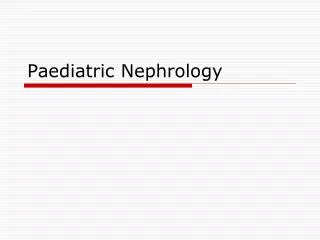
Urine microscopy • RBCs • Infection, glomerular disease, malignacy • WBCs • Infection, glomerulopnehritis, malignancy, TB, intersitial nephritis, inflammation • Crystals • Seen with stones, cystinuria, gout • Casts • Hyaline casts (clear and colourless) • Seen with exercise, fever, concentrated urine (often in normal subjects) • Red cell casts • Seen in GN, vasculitis, malignant hypertension • White cell casts • Pyelonephritis, proliferative glomerulonephritis • Epithelial casts • Acute tubular necrosis, acute glomerulonephritis • Granular casts • GN, diabetic nephropathy, amyloidosis, intersitial nephritis
RBC WBC Granular Hyaline Casts
24 Hour urine collection • Void , discard urine, note time • Collect all urine 24 hours • Exactly 24 hours later empty bladder and collect urine • Provide serum sample for creatinine • Measure volume, protein, creatinine, sodium
Protein • Excrete 80-150mg/day • Minimum concentration for dipstick detection 20-30 mg/dl • Glomerular disease • suspected >1g/24 hours • certain > 3g/24 hours • Tubular disease - <3g/24 hours • Overflow – abnormal immunoglobins • Myeloma – bence jones protein
Urinary Albumin:Creatinine ratio • Proteinuria is a measure of renal disease • 24 urinary protein estimation is a pain • 3 samples over 2 months • Spot microalbuminuria test is subject to variability • Comparing with creatinine improves its accuracy • Normal <2.5mg/mmol • <30 ‘acceptable’ • 30-70 warrants a retest • >70mg/mmol NICE suggests referral to nephrology
Case 3 • 60 yr old woman • Mild renal impairment (eGFR 59) • Hypertension • 4 cm exophytic, enhancing, solid, left renal mass • Has a partial nephrectomy • Seen for follow-up 3 months later
Case 3 • She wants to know how much kidney function has she got left? • Biochemical tests • Nuclear medicine tests
Case 3 • Biochemical tests • Assess overall function • Various methods for refining assessment, but none perfect • Radiological tests • Can assess overall and relative function • Can give additional information eg Scars, drainage
Blood tests • Urea • Varies with protein and catabolic states • Freely filtered • Reabsorbed at variable rate dependant on water reabsorption • Not a reliable indicator of function • Creatinine • Freely filtered and secreted into distal tubule • Varies with muscle mass
Serum creatinine not sensitive indicator in early renal impairment • • Need GFR to fall below 60-80ml/min before there is rise in Serum Cr • • An abnormal creatinine indicates a loss of 50% of renal function
MCQ • MDRD formula is not valid for children • The Cockcroft & Gault formula is an alternative to MDRD for calculating eGFR • MDRD formula uses 3 variables to calculate eGFR • Your path lab should use the same formula as the online MDRD calculator • eGFR is accurate an reproducible in acute renal failure
MCQ • MDRD formula is not valid for children • The Cockcroft & Gault formula is an alternative to MDRD for calculating eGFR • MDRD formula uses 3 variables to calculate eGFR • Your path lab should use the same formula as the online MDRD calculator • eGFR is accurate an reproducible in acute renal failure
Formulae • Used to improve on serum creatinine, but all suffer from its limitations • Estimated creatinine clearance • Cockcroft-Gault • Estimated GFR • MDRD (4 and 6 variable) • CKD-EPI • Mayo
Cockcroft and Gault formula • Creatinine clearance = (140-age) x body weight in Kg 72 x serum creatinine in mg/dL • Multiply by 0.85 for women • May be more accurate than timed urine collections • Assumptions: • Lean body weight (hence in obesity, will overestimate) • Volume distribution and Creatinine production is in steady state (hence will overestimate in low protein diet)
MDRD • 4 variable (Creatinine, Age, Gender, Race) • 6 variable (Albumin, Urea) • eGFR=32788 x [creatinine in μmol/L]-1.154 x Age-0.203 • x 1.212 if black • x 0.742 if female • Should be automatically reported by your lab every time serum creatinine is checked. • Be aware of its limitations
Limitations of MDRD • It is only an estimate, significant error is possible. Likely to be inaccurate in extremes of body type • malnourished, • amputees, • It is not valid in pregnant women • Some racial minorities may not fit the MDRD equation well. Originally validated for US white and black patients. • Not so good near normal: The MDRD equation tends to underestimate normal or near-normal function. Routine reporting of eGFR values >90 is not recommended.
Limitations of MDRD • Creatinine level must be stable: eGFR calculations assume that the level of creatinine in the blood is stable over days or longer. They are not valid if it is changing. • The MDRD equation is not valid for under-18s. Use the Counahan-Barrat method for children • Different equations: from April 2006 in the UK, local laboratories should calculate eGFR on all samples sent for creatinine measurement. The equation they use will take into account local variations in accuracy of creatinine assays, so eGFR values obtained in this way should be a little more accurate than those generated by any of the online calculators
CKD-EPI • eGFR = 141 x min([creat]/κ,1)α x max([creat]/κ,1)-1.209 x 0.993Age x 1.018 [if female] x 1.159 [if black] • κ = 0.7 if female.κ = 0.9 if male. • α = -0.329 if femaleα = -0.411 if male • min = the minimum of Scr/κ or 1max = the maximum of Scr/κ or 1 • Probably more accurate than MDRD • Certainly better if GFR > 60mls/min • New (2009) and MDRD remains the NICE approved formula
EMQ A. Inulin clearance E. Cockcroft and Gault B. Cr51 EDTA F. Tc99m DMSA C. Tc99m DTPA G. 24hr urinary creatinine clearance D. MDRD 6 variable H. eGFR Which of the above: • Could be used to assess GFR and renal drainage at the same time • Is the gold standard for assessment of glomerular filtration rate • Is not an assessment of overall renal function
EMQ A. Inulin clearance E. Cockcroft and Gault B. Cr51 EDTA F. Tc99m DMSA C. Tc99m DTPA G. 24hr urinary creatinine clearance D. MDRD 6 variable H. eGFR Which of the above: • Could be used to assess GFR and renal drainage at the same time B • Is the gold standard for assessment of glomerular filtration rate A • Is not an assessment of overall renal function F
Creatinine Clearance • 24 urinary creatinine measurement allows calculation of creatinine clearance • Clearance = UV/P (U = urine concentration, V = flow rate, P = plasma concentration) • Usually adjusted for body surface area • Normal >120ml/min/1.73m2 • Creatinine clearance is 20% higher than GFR due to tubular excretion of creatinine
Glomerular filtration rate • GFR determined by using substance • Neither metabolised nor synthesised • Secreted into plasma at constant rate • freely filtered at glomerulus • Neither secreted or absorbed further down nephron • Inulin clearance is the gold standard for GFR measurement • Polysaccharide • Filtrated (not reabsorbed, secreted or metabolised by the kidney) • Continuous infusion • Measure Inulin in urine and blood until steady state reached • Expensive, time consuming, impractical
Isotopic GFR • Cr51 EDTA (closest in clearance pattern to inulin) • Tc99m DTPA (short half life, but can do a renogram at the same time) • Blood sample for background count • Known dose of radiopharmaceutical given • Time for equilibration • Samples at 2,3 and 4 hours • Volume of distribution worked out • Rate of clearance calculated by slope intercept
Renography • A study of the uptake, transit and elimination by the kidney of an intravenous dose of a radionucleotide • Gives information on drainage and relative function • Limited anatomical information • Use of diuretic improves discrimination between obstructed and non-obstructed patterns • Tc99m MAG-3 • I131 Hippuran • T99m DTPA • See nephrology 1 for renograms in detail
DMSA scan • Dimercaptosuccinic acid • Bound in proximal tubules • Excellent imaging of functioning areas of cortex • IV injection with imaging 3 hours later • Multiple views to allow better visualisation • Can calculate relative function • Look for areas of poor function ie scars • May need to wait 6 months after last insult for areas to recover