Radiation Protection
Radiation Protection. Robert L. Metzger, Ph.D. 1. Sources of Exposure to Ionizing Radiation Naturally Occurring Radiation Sources.

Radiation Protection
E N D
Presentation Transcript
Radiation Protection Robert L. Metzger, Ph.D.
1. Sources of Exposure to Ionizing RadiationNaturally Occurring Radiation Sources • Annual average total effective dose from exposure to ionizing radiation in USA is approximately 3.6 mSv or 360 mrem [National Council on Radiation Protection and Measurement (NCRP)] • 3 mSv or 300 mrem (80%) is from naturally occurring sources • Radon • Internal radiation • Terrestrial radioactivity • Cosmic radiation c.f. Bushberg, et al. The Essential Physics of Medical Imaging, 2nd ed., p. 748.
Radon Biggest contributor to natural background (2 mSv or 200 mrem/year) Radon (Rn-222) is a radioactive gas formed during the decay of radium Radium is a decay product of uranium found in the soil and has a half-life of 1620 years Radon is an alpha emitter with a half-life of approx. 4 days 1. Sources of Exposure to Ionizing RadiationNaturally Occurring Radiation Sources c.f. Bushberg, et al. The Essential Physics of Medical Imaging, 2nd ed., p. 748.
Radon The progeny of radon are also radioactive, attach to aerosols and are deposited in the lungs Bronchial mucosa is irradiated inducing bronchogenic cancer Average concentration of radon outdoors is 4-8 Bq/m3 (0.2-0.4 pCi/L) Indoors is 40 Bq/m3 (1 pCi/L) EPA Remedial action recommended in excess of 160 Bq/m3 (4 pCi/L) 1. Sources of Exposure to Ionizing RadiationNaturally Occurring Radiation Sources
Internal Radiation Second largest source of natural background radiation (0.4 mSv or 40 mrem/year) Ingestion of food and water containing primordial radionuclides K-40 is most significant Skeletal muscle has the highest concentration of potassium in the body 1. Sources of Exposure to Ionizing RadiationNaturally Occurring Radiation Sources c.f. Bushberg, et al. The Essential Physics of Medical Imaging, 2nd ed., p. 748.
Terrestrial or External Radiation Terrestrial radioactive materials that have been present on earth since its formation are called primordial radionuclides External radiation exposure, inhalation, ingestion 0.28 mSv or 28 mrem/year ( 0.3 mSv or 30 mrem/year) 1. Sources of Exposure to Ionizing RadiationNaturally Occurring Radiation Sources c.f. Bushberg, et al. The Essential Physics of Medical Imaging, 2nd ed., p. 748.
Cosmic Radiation Cosmic rays are energetic protons and alpha particles which originate in galaxies Most cosmic rays interact with the atmosphere, with fewer than 0.05% reaching sea level 0.27 mSv or 27 mrem/year ( 0.3 mSv or 30 mrem/year) 1. Sources of Exposure to Ionizing RadiationNaturally Occurring Radiation Sources c.f. Bushberg, et al. The Essential Physics of Medical Imaging, 2nd ed., p. 748.
Cosmic Radiation Exposures increase with altitude approx. doubling every 1500 m as there is less atmosphere to attenuate the cosmic radiation Leadville, Colorado at 3200 m, 1.25 mSv/year More at poles than equator 1. Sources of Exposure to Ionizing RadiationNaturally Occurring Radiation Sources c.f. Bushberg, et al. The Essential Physics of Medical Imaging, 2nd ed., p. 748.
Cosmic Radiation Air travel can add to individual’s cosmic exposure Airline crews and frequent fliers receive an additional 1 mSv 5 hour transcontinental flight will result in an equivalent dose of 25 mSv or 2.5 mrem Apollo astronauts – 2.75 mSv or 275 mrem during a lunar mission 1. Sources of Exposure to Ionizing RadiationNaturally Occurring Radiation Sources c.f. Bushberg, et al. The Essential Physics of Medical Imaging, 2nd ed., p. 748.
Annual Dose - Other 1% • Occupational Dose – 0.3% • Fallout - <0.3% • Nuclear Fuel Cycle – 0.1% • Miscellaneous – 0.1% • Natural Sources Account for 82% of total annual dose with only 18% coming from man made sources.
Natural Sources of Radiation • The variation of dose from the cosmic and terrestrial radiation is large depending on the area of the country (see handout). • High altitude areas have higher cosmic radiation levels (e.g. Denver, Flagstaff) • Areas that are heavily mineralized have higher terrestrial radiation levels. • Radon levels also vary significantly, but vary from home to home rather than whole geographic areas (e.g. Watras house) • The overall variation in cosmic and terrestrial radiations exceed 100 mrem per year and affect everyone in a city/area (e.g. Denver) • Cancer incidence does not follow the background radiation levels at all.
Sources of Exposure to Ionizing RadiationTechnology Based Radiation Sources • 60 mrem or 0.6 mSv • CT and fluoroscopy • are highest contributors • to medical • x-rays c.f. Bushberg, et al. The Essential Physics of Medical Imaging, 2nd ed., p. 744.
1. Occupational Exposures • 1 mSv for diagnostic radiology is lower than expected because it includes • personnel who receive very small occupational exposures • 15 mSv or more are typical of special procedures utilizing fluoroscopy and cine c.f. Bushberg, et al. The Essential Physics of Medical Imaging, 2nd ed., p. 745.
1. Collective Effective Dose Equivalent • The product of the average effective dose equivalent and the size of the exposed population is the collective effective dose equivalent • Expressed in person-sieverts (person-Sv or person-rem) [not used much anymore] c.f. Bushberg, et al. The Essential Physics of Medical Imaging, 2nd ed., p. 746.
1. Genetically Significant Dose (GSD) • The genetically significant equivalent dose (GSD) is a dose parameter that is an index of potential genetic damage • The GSD is defined as that equivalent dose that, if received by every member of the population, would be expected to produce the same genetic injury to the population as do the actual doses received by the irradiated individuals • GSD is determined by taking the equivalent dose to the gonads of each exposed individual and estimating the number of children expected for a person of that age and sex
1. Genetically Significant Dose (GSD) c.f. Bushberg, et al. The Essential Physics of Medical Imaging, 2nd ed., p. 747.
1. Summary • The average annual effective dose equivalent to the US population from all radiation sources is 3.6 mSv/year or 360 mrem/year • 3 mSv/year – naturally occurring sources • Radon – 2 mSv • 0.6 mSv/year – technologically enhanced sources • Medical x-rays – 0.39 mSv or 39 mrem, • Nuclear Medicine – 0.14 mSv or 14 mrem • Data is from mid – 80s. Current estimates are higher due to the increased use of CT. Recent estimates are 385 mrem with 85 mrem due to medical.
1. Summary • Collective effective dose equivalent (person-Sv or person-rem) • Product of the average effective dose equivalent and the size of the exposed population (No longer used commonly) • GSD (mSv or mrem) • Used to express genetic risk to the whole population from a source of radiation exposure • GSD from diagnostic x-rays is 0.2 mSv or 20 mrem • GSD from nuclear medicine is 0.02 mSv or 2 mrem
Raphex 2002 General Question • G87. The annual average natural background radiation dose to members of the public in the United States, excluding radon, is approximately ________ mrem. • A. 10 • B. 50 • C. 100 • D. 200 • E. 400
Question • 1. The Genetically significant dose (GSD) for diagnostic x-rays and nuclear medicine in the US is: • A. 2 mSv and 0.20 mSv • B. 0.20 mSv and 2 mSv • C. 0.02 mSv and 0.20 mSv • D. 0.20 mSv and 0.02 mSv
2. Personnel DosimetryFilm Badges • A film pack (A) consists of a black envelope (B) containing film (C) placed inside a special plastic film holder (D) • Using metal filters typically lead (G), copper (H) and aluminum (I), the relative optical densities of the film underneath the filters can be used to identify the general energy range of the radiation and allow for the conversion of the film dose to tissue dose • Open window (J) where film is not covered by a filter or plastic and is used to detect medium and high-energy beta radiation c.f. Bushberg, et al. The Essential Physics of Medical Imaging, 2nd ed., p. 749.
2. Personnel DosimetryFilm Badges • Most film badges can record doses from about 100 mGy to 15 Gy (10 mrad to 1500 rad) for photons and from 500 mGy to 10 Gy (50 mrad to 1,000 rad) for beta radiation • The dosimetry report lists the “shallow” equivalent dose, corresponding to the skin dose, and the “deep” equivalent dose, corresponding to penetrating radiation • Generally placed at waist level or shirt-pocket level • For fluoroscopy, placed at collar level outside the lead apron to measure radiation dose to thyroid and lens of eye • Pregnant radiation workers typically wear a second badge at waist level (behind the lead apron, if used) to assess the fetal dose • Excessive moisture or heat will damage film inside badge
2. Personnel DosimetryThermoluminescent (TLD) Dosimeters • TLD is a dosimeter in which consists of a scintillator in which electrons become trapped in excited states after interactions with ionizing radiation • If the scintillator is later heated, the electrons can then fall to their ground state with the emission of light • Thermoluminescent (TL) means emitting light when heated • The amount of light emitted by the TLD is proportional to the amount of energy absorbed by the TLD • After TLD has been read, it may be baked in an oven and reused
2. Personnel DosimetryThermoluminescent (TLD) Dosimeters • Lithium Fluoride (LiF) is one of the most useful TLD materials • LiF TLDs have a wide dose response range of 10 mSv to 103 mSv (1 mrem to 105 rem) • Used in nuclear medicine to record extremity exposures
2. Personnel DosimetryOptically Stimulated Luminescent (OSL) Dosimeters • The principle of OSL is similar to TLDs except that the light emission is stimulated by a laser light instead of heat • Crystalline aluminum oxide activated with carbon (Al2O3:C) is commonly used • Broad dose response range like TLDs • They can be reread several times
2. Personnel DosimetryPocket Dosimeters • Major disadvantage to film and TLD dosimeters is that the accumulated exposure is not immediately indicated • Pocket dosimeters measure radiation exposure, which can be read instantaneously • Can measure exposures from 0 to 200 mR or 0 to 5 R • Analog or digital c.f. Bushberg, et al. The Essential Physics of Medical Imaging, 2nd ed., p. 752.
2. Summary c.f. Bushberg, et al. The Essential Physics of Medical Imaging, 2nd ed., p. 753.
Raphex 2002 General Questions • G95. Film badges: • A. Can measure only the total dose of radiation, but cannot distinguish between low and high energy x-rays. • B. Can measure exposures of 1 mR. • C. Are insensitive to heat. • D. Use the optical density of the film to measure dose.
3. Radiation Detection Equipment In Radiation Safety • Geiger-Mueller Survey Instruments • Measurements are in counts per minute (cpm) • Surveys radioactive contamination in nuclear medicine • Are extremely sensitive to charged particulate radiations with sufficient energy to penetrate the survey meter window • Are relatively insensitive to x- and gamma radiations • Portable Ionization Chambers • Used when accurate measurements of radiation exposure are required, measurement of x-ray machine outputs • Measure 1 mR/hr to 500 R/hr
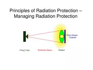
4. Radiation Protection and Exposure Control • There are four principal methods by which radiation exposures to persons can be minimized: time, distance, shielding and contamination control • Time • reducing time spend near a radiation source • Distance • inverse square law • For diagnostic x-rays, a good rule of thumb is that at 1 m from a patient at 90 degrees to the incident beam, the radiation intensity is 0.1% to 0.15% (0.001 to 0.0015) of the intensity of the beam incident upon the patient for a 400 cm2 area field area • The NCRP recommends that personnel should stand at least 2 m from the x-ray tube and the patient and behind a shielded barrier or out of the room, whenever possible
4. Radiation Protection and Exposure ControlShielding • Shielding is used to reduce exposure to patients, staff and the public • Shielding against primary (focal spot), scattered (patient) and leakage (x-ray tube housing, limited to 100 mR/hr at 1 m from housing) radiation
Shielding – NCRP 49 vs. 147 • The Bushberg X-Ray shielding section is based on NCRP 49 which is obsolete and out of print. Bushberg notes that it is badly out of date. • NCRP 147 replaced NCRP 49 in 2005. It should be used for all shielding calculations. • NCRP 147 uses a different methodology to calculate the shielding values and uses much more realistic values for occupancy, tube kVps, weekly mAs, and film/screen speeds. • It eliminates the gross overshielding resulting from the use of NCRP 49 with the lowered non-occupational dose limits. • NCRP 147 also provides shielding methodologies for CT and othr modalities.
4. Radiation Protection and Exposure ControlShielding • Shielding calculations depend on: • radiation exposure level (mR/week) depends on techniques and patient load • workload (amount of x-rays produced per week), W (mA.min/week) • use factor, U, indicates the fraction of time during which the radiation under consideration is directed at a particular barrier • a wall that intercepts the primary beam is called a primary barrier and is assigned a use factor according to typical room use • U ranges between 0 and 1, secondary barriers have a use factor of 1
4. Radiation Protection and Exposure ControlShielding • Shielding calculations depend on: • occupancy factor, T, indicates the fraction of time during a week that a single individual might spend in an adjacent area • T = 1 for full occupancy (work areas, offices etc.) • T = 1/5 for partial occupancy (corridors, rest rooms etc.) • T = 1/16 for occasional occupancy (waiting rooms, toilets, etc.) • T = 1/40th for landscaping, etc. • Distance, d, measured from source of radiation to the area to be protected
4. Radiation Protection and Exposure ControlShielding • Shielding calculations determine the thickness of an attenuating material required to reduce radiation exposure to acceptable levels • 1 mSv/year or 100 mrem/year (2 mR/week) for non-occupational personnel (members of public and non-radiation workers) • 0.1 or 10 mR/week for controlled areas (pregnant worker limit)
4. Radiation Protection and Exposure ControlShielding • Lead usually used for shielding and specified as weight per square foot (lb/ft2). Typically 2 lb/ft2 (0.8 mm or 1/32th inch) or 4 lb/ft2 (1.6 mm or 1/16th inch) is sufficient for diagnostic radiology • Calculated using HVL and TVL of the material [(1/2)n – reduction in beam intensity, n is HVL] • Shielding material used from base of floor to a height of 7 feet • Acrylic, leaded glass, gypsum drywall, steel are other materials used besides lead for shielding
4. Radiation Protection and Exposure Control • CT scanner shielding (Use NCRP 147 with web based scatter values) • Personnel protection in Dx Radiology (lead aprons, thyroid shields etc., pg. 771 of Bushberg) • Shielding in nuclear medicine • Shielding in PET (Beware!) Undershielding in some clinics have led to high technologist and non-occupational doses. PET shielding guide from AAPM is not published as yet.
4. Radiation Protection and Exposure ControlProtection of the Patient in Medical X-ray Imaging • Tube Voltage and Beam Filtration • Achieve an optimal balance between image quality and dose to the patient • Patient exposure can be reduced by using a higher kVp ad lower mAs • Increasing kVp increases transmission (less absorption) of x-rays through the patient • Even though mR/mAs increases as kVp increases, an accompanying reduction in mAs will decrease the incident exposure to the patient • Contrast will decrease due to higher effective energy of the x-ray beam
4. Radiation Protection and Exposure ControlProtection of the Patient in Medical X-ray Imaging • Tube Voltage and Beam Filtration • Filtration of the polychromatic x-ray energy spectrum can significantly reduce exposure by selectively attenuating the low-energy x-rays in the beam • As the tube filtration increases, the beam becomes hardened (effective energy increases) and dose to patient decreases because fewer low-energy photons are in the incident beam • The amount of filtration that can be added is limited by the increased demands on tube loading to offset reduction in tube output, and the decreased contrast due to excessive beam hardening • Quality of x-ray beam is assessed by measuring the HVL
Depth Dose • Recall Dose = Energy absorbed per gram. • For soft radiations, the dose decreases dramatically with depth as the patient’s body attenuates the beam. • The radiation dose at a given depth is the depth dose (rad). • The exposure at skin entrance (ESE) is the Roentgen exposure at the point where the radiation enters the body.
4. Radiation Protection and Exposure ControlProtection of the Patient in Medical X-ray Imaging • Field Area, Organ Shielding and Geometry • Reducing field size limits the patient volume exposed to primary beam, reduces the amount of scatter and thus radiation dose to adjacent organs (scatter being reduced improves image contrast) • Gonadal shielding can be used to protect the gonads from primary radiation when the shadow of the shield does not interfere with the anatomy under investigation • Increasing source-to-object distance (SOD) and source-to-image distance (SID) helps reduce dose (patient volume exposed decreased due to reduced beam divergence) • For fixed SID (C-arm fluoro system), patient dose is reduced by increasing the SOD as much as possible • A minimum patient to focal spot distance of 20 cm is required
4. Radiation Protection and Exposure ControlProtection of the Patient in Medical X-ray Imaging • X-Ray Image Receptors • The speed of the image receptor determines the number of x-ray photons and thus the patient dose necessary to achieve an appropriate signal level • Higher speed system requires less exposure to produce the same optical density and thus reduces dose to patient • Either a faster screen (reduced spatial resolution) or faster film (increased quantum mottle) will reduce the incident exposure to the patient
4. Radiation Protection and Exposure ControlProtection of the Patient in Medical X-ray Imaging • X-Ray Image Receptors • Computed Radiography (CR) devices have a wide dynamic range so they compensate to some degree for under- and overexposure and can reduce retakes • CR roughly equivalent to 200 speed screen-film systems • Techniques for extremities with CR devices should be used at higher exposure levels while exposures for pediatric patients should be used at increased speed (e.g. 400 speed) to reduce dose
4. Radiation Protection and Exposure ControlProtection of the Patient in Medical X-ray Imaging • Computed Tomography (CT) • Reduce mAs and perhaps kVp for thinner and pediatric patients • Pediatric protocols required in AZ. • Modern MSCT scanners – dose modulation, mA changes with patient size c.f. Bushberg, et al. The Essential Physics of Medical Imaging, 2nd ed., p. 779.
4. Radiation Protection and Exposure ControlProtection of the Patient in Medical X-ray Imaging • Miscellaneous Considerations • Careful identification of patients • Determination of pregnancy status • Eliminate screening exams that only rarely detect pathology • “yearly” dental exams may not be appropriate for all patients • Use of high speed dental film reduces dose • “yearly” screening mammography exams not appropriate for women younger than 35 to 40 years old • Technique errors and high repeat rates can be avoided by posting technique charts and using phototiming • Good quality control program to eliminate equipment and processor problems
4. Summary • Time, distance and shielding used to protect persons from radiation exposure • Shielding calculations depend on mR/week, workload, use factor, occupancy factor and distance from x-ray source • Typically 2 or 4 lb/ft2 lead is sufficient for shielding in diagnostic radiology • Calculated using HVL and TVL of the material [(1/2)n – reduction in beam intensity, n is HVL] • Protect patient by adjusting kVp, mAs, filtration, field size, geometry and using organ shielding, using faster film-screen systems, eliminate screening chest and yearly dental exams