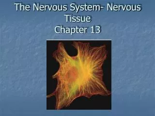
CHAPTER 13 Nervous Tissue
CHAPTER 13 Nervous Tissue. COMMON COURSE OBJECTIVES: Functions of the nervous system Organization of the nervous system Nerve tissue and nerve cell types Structure of a typical neuron Structure of a chemical synapse. Nervous Tissue Histology. Composed of:

CHAPTER 13 Nervous Tissue
E N D
Presentation Transcript
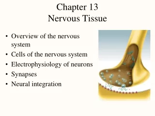
CHAPTER 13Nervous Tissue COMMON COURSE OBJECTIVES: Functions of the nervous system Organization of the nervous system Nerve tissue and nerve cell types Structure of a typical neuron Structure of a chemical synapse
Nervous Tissue Histology • Composed of: • Neurons true conducting cells in nervous tissue • Neuroglial (supporting) cells -Astrocytes -Schwann cells -Oligodendrocytes -Satellite cells -Microglia -Ependymal
The NERVOUS SYSTEM • Defined: like the CPU of a computer, the nervous system is the master controlling system of the body. It is designed to constantly and rapidly adjust and respond to stimuli the body receives. It includes the brain, cranial nerves, spinal cord, and associated peripheral nerves.
Properties of Neurons • Excitability (irritability): ability to respond to environmental changes or stimuli. • Conductivity: respond to stimuli by initiating electrical signals that travel quickly to other cells at distant locations. • Secretion: Upon arrival of the impulse at a distant location the neuron usually secretes a chemical neurotransmitter at a synapse that crosses the synaptic gap and stimulates the next cell.
Functional Classes of Neurons • Sensory (afferent) neurons – afferent neurons are specialized to detect stimuli and transmit the information to CNS. They begin in any organ in the body, but end in the brain or spinal cord. • Interneuron (association neurons): lie entirely in the CNS. They receive signals from many different neurons and perform an integrative function “decision making” to respond to the different stimuli. • Motor (efferent) neurons – efferent neurons transmit the appropriate response from the interneuron to an end organ (muscle and gland cells) to carry out the body’s response to the stimuli.
Functional Classification of Neurons • Based on the direction of conduction • Sensory or afferent conduct toward the CNS • Motor or efferent conduct away from the CNS • Interneuron interposed between sensory and motor
Organization of the Nervous System Two main divisions: • The Central Nervous System (CNS) - Consists of the brain and spinal cord with tracts and nuclei Nucleus = a collection of nerve cell bodies in the CNS. Tract = bundle of nerve fibers within the CNS • The Peripheral Nervous System (PNS) -Consists of ganglia, cranial nerves, spinal nerves and peripheral receptors Ganglia = a collection of nerve cell bodies in the PNS Nerve = bundle of nerve fibers in the PNS
Peripheral Nervous System (PNS) • Composed of cranial nerves and spinal nerves and their branches, ganglia and sensory receptors. • PNS is subdivided into sensory and motor divisions: • somatic nervous system (SNS) • autonomic nervous system (ANS) and the • enteric nervous system (ENS)
Sensory or Afferent Division • Somatic sensory = senses touch, pressure, pain, temperature, vibration and proprioception in skin, body wall and limbs. • Visceral sensory = Autonomic sensory division-sensesstretch, pain, temperature, chemical changes and irritation in viscera; nausea and hunger.
Motor or Efferent Division • Somatic motor -motor control to all skeletal muscles except pharyngeal muscles. • Visceral Motor = Autonomic Nervous System – Four “F’s” -Sensory receptors convey information from visceral organs (e.g. heart, lungs, intestines, etc.) to the CNS for integration and interpretation. A motor response is initiated that conducts impulses from CNS to smooth muscle, cardiac muscle and/or glands for appropriate response
Autonomic Nervous System • Two divisions of ANS (4 “F’s”) • Sympathetic division – Fight or Flight • Parasympathetic division – Food or Sex
Neurons Nerve cell proper Cell body (soma) Dendrites - TO Axons -FROM
Structural Classification of Neurons • Neurons may be: Multipolar, Bipolar or Unipolar • Determined by the number of processes attached to the cell body
Neurons • Most (99%) neurons in the body are multipolar. • Bipolar neurons are rare and occur in special sense organs of ear, nose and eye. • Unipolar neurons begin as bipolar but processes fuse into one. They are primarily sensory neurons.
Neuroglia cells • Found in CNS and PNS • Perform a supporting function for neurons • CNSPNS • Oligodendroglialcytes Schwann cells • Astrocytes Satellite cells • Ependymal cells • Microglia
Oligodendrogliocytes -CNS • Form myelin sheath in CNS • Fewer branches than astrocytes
Myelin • Insulating layer around a nerve • Formed by oligodendrocytes in CNS and Schwann cells in PNS • Composed of a lipoprotein with phospholipids, glycolipids and cholesterol. • Myelination is process of myelin formation
Astrocytes - CNS Star shaped Blood brain barrier Most numerous
Ependymal cells - CNS • Epithelial cells that line ventricles and central cavities of brain and spinal cord-secrete CSF • Ciliated to circulate CSF
Microglia - CNS • Thorny bushes in appearance and the smallest glia • Phagocytic function in CNS • Originate from monocytes
Schwann cells- PNS • Form myelin sheath around peripheral axons • Look like jelly roll with neurolemma cover • Node of Ranvier separate each Schwann cell
Satellite cells -PNS • Surround neuron cell bodies within ganglia
Synaptic ending • Synapse – site where two nerves communicate with each other. • Presynaptic neuron – neuron that is conducting information toward the next neuron • Postsynaptic neuron – transmits information away from synapse • Most synaptic communication is via chemical messengers (e.g. acetylcholine, serotonin, norepinephrine, dopamine, endorphins, GABA, glycine, glutamic acid, etc.)
Types of synapses • Axodendritic = axon to dendrite • Axosomatic = axon to cell body • Axoaxonic = axon to axon • Dendrodendritic = dendrite to dendrite • Dendrosomatic = dendrite to cell body