Chapter 18
Chapter 18. The Genetics of Viruses and Bacteria. 0.5 m. Figure 18.1 T4 bacteriophage infecting an E. coli cell. Virus. Bacterium. Animal cell. Animal cell nucleus. 0.25 m. Figure 18.2 Comparing the size of a virus, a bacterium, and an animal cell. THE GENETICS OF VIRUSES.

Chapter 18
E N D
Presentation Transcript
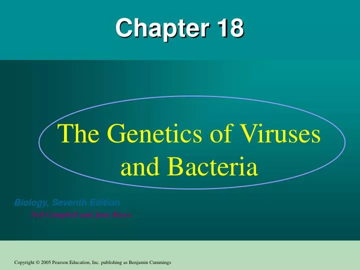
Chapter 18 The Genetics of Viruses and Bacteria
0.5 m Figure 18.1 T4 bacteriophage infecting an E. coli cell
Virus Bacterium Animalcell Animal cell nucleus 0.25 m Figure 18.2 Comparing the size of a virus, a bacterium, and an animal cell
THE GENETICS OF VIRUSES • How viruses were discovered? • The story begins in 1883 with Adolf Mayer from Germany who was studying the cause of tobacco mosaic disease. • Mayer discovered that the disease was contagious • He tried to see any thing in the contagious sap extracted form infected plants but could not seen any thing. • He concluded that the disease is caused by an unusually small organism
THE GENETICS OF VIRUSES • Later in 1897 Martinus Beijerinck discovered that the infectious agent can reproduce but only in the agent that infects but NOT in the nutrient media unlike bacteria. In addition the agent was not inactivated by alcohol which inactivate bacteria. • 1n 1935 the American Scientist Wendell Stanley crystallized the infectious particle that is now know as tobacco mosaic virus (TMV). • Later with the aid of the electron microscopy, the virus was seen.
So what is the virus? • A virus is a genome enclosed in a protective coat • The tiniest virus is only 20 nm in diameter that is smaller than a ribosome. • The largest virus can barely be resolved by light microscope. • Viruses are infectious particles consisting of nucleic acids enclosed in a protein coat and some times a membranous envelope.
Figure 18.3 Infection by tobacco mosaic virus (TMV) Normal Infected
Structure of Viruses • Viruses are called DNA or RNA viruses based on their genetic material which could consist of; • double stranded DNA • Single stranded DNA • Double stranded RNA • Single stranded RNA • Smallest viruses have only 4 genes while the largest have several hundred genes.
Capsids and envelopes • The protein shell that encloses the viral genome is called the capsid which is made large number of protein subunits called capsomeres. • Tobacco mosaic virus has a structure that contains over a thousand molecules of a single type of protein (helical) • Adeno virus that causes respiratory infection in animals made of 252 identical protein molecules (polyhydral). • Influenza virus has a viral envelopes contain proteins, glycoproteins and phospholipids • The most complex capsids are found among viruses that infect bacteria (bacteriophages). There are 7 types of bacteriophages that infect E. coli called T1 –T7
Capsomereof capsid Membranousenvelope RNA Capsomere DNA Head Capsid Tail sheath DNA RNA Tail fiber Glycoprotein Glycoprotein 80 225 nm 18 250 mm 80–200 nm (diameter) 70–90 nm (diameter) 50 nm 20 nm 50 nm 50 nm (d) Bacteriophage T4 (a) Tobacco mosaic virus (b) Adenoviruses (c) Influenza viruses Figure 18.4 Viral structure
Viruses can reproduce only within a host cell • Viruses are obligate intracellular parasites that is they reproduce only within a host cell. • Viruses have no enzymes for metabolism and have no ribosomes or other equipement for making their own proteins. • Each virus can infect only a limited range of hosts called the host range. • Viruses identify their hosts by a lock and key mechanism. However some viruses have wider range than others such as swine flue virus can infect both humans and hogs while rabies virus can infect a number of mammalian species including raccoons, skunks, dogs and humans. • Viruses of eukaryotes are usually tissue specific such as human cold virus that infects upper respiratory tract or AIDS virus that attaches to CD4 cells of the immune system.
How does a viral infection occur (Figure 18-5) • A viral infection begins when a virus genome finds its way to a host cell by the specific mechanism of injection used by the virus. • Once inside, the viral genome can commandeer its host, reprogram the cell to copy the viral nucleic acid and manufacture viral proteins • Most viruses use DNA polymerase of the host cell to synthesize new genomes along the template provided by viral DNA. • With regard to RNA viruses they use special virus-encode polymerase and use RNA as template. • The host provide all the resources for nucleic acid synthesis such as nucleotides (N), enzymes, ribosomes, tRNAs, amino acids, ATP and other components needed for making proteins as dictated by the viral genes. • After the production of capsid proteins and the replication of viral DNA their assembly of new viruses is spontaneous. • The cycle completes after that hundreds or thousands emerging from the infected cell causing the death of the cell and infecting hundreds or thousands of other cells.
VIRUS Entry into cell and uncoating of DNA DNA Capsid Transcription Replication HOST CELL Viral DNA mRNA Viral DNA Capsid proteins Self-assembly of new virus particles and their exit from cell Figure 18.5 A simplified viral reproductive cycle
Attachment. The T4 phage usesits tail fibers to bind to specificreceptor sites on the outer surface of an E. coli cell. Entry of phage DNA and degradation of host DNA.The sheath of the tail contracts,injecting the phage DNA intothe cell and leaving an emptycapsid outside. The cell’sDNA is hydrolyzed. 1 2 4 3 5 Release.The phage directs productionof an enzyme that damages the bacterialcell wall, allowing fluid to enter. The cellswells and finally bursts, releasing 100 to 200 phage particles. Phage assembly Synthesis of viral genomes and proteins. The phage DNAdirects production of phageproteins and copies of the phagegenome by host enzymes, usingcomponents within the cell. Assembly. Three separate sets of proteinsself-assemble to form phage heads, tails,and tail fibers. The phage genome ispackaged inside the capsid as the head forms. Head Tail fibers Tails Figure 18.6 The lytic cycle of phage T4, a virulent phage
Phage DNA The phage attaches to a host cell and injects its DNA. Many cell divisions produce a large population of bacteria infected with the prophage. Phage DNA circularizes Phage Occasionally, a prophage exits the bacterial chromosome, initiating a lytic cycle. Bacterial chromosome Lytic cycle Lysogenic cycle Certain factors determine whether The bacterium reproduces normally, copying the prophage and transmitting it to daughter cells. The cell lyses, releasing phages. Lytic cycle is induced Lysogenic cycle is entered Prophage or New phage DNA and proteins are synthesized and assembled into phages. Phage DNA integrates into the bacterial chromosome,becoming a prophage. Figure 18.7 The lytic and lysogenic cycles of phage , a temperate phage
With this mechanism how come that bacteriophage have not exterminated all the bacteria ? • Bacteria are not defense less. • Natural selection favors bacterial mutants with receptor sites that are no longer receptive for a particular bacteriophage. • When the virus inters several enzymes might break it down, such enzymes are called restriction endonucleasis. • Bacterial DNA is chemically modified so that it can not be destroyed by these restriction enzymes. • Some phages can live inside the cell without lysing it instead coexist in what is called lysogenic cycle.
The Lysogenic cycle • Unlike the lytic cycle that kills the cell, lysogenic cycle replicates the phage genome without destroying the host. • The phages that are capable of using both modes are called temperate phages. • An example of a temperate phage is called lamba (λ) and Figure 18-5 shows the Lysogenic and lytic reproductive cycles of phage λ • During the Lysogenic cycle the viral genome behaves differently, the λ DNA molecule is incorporated (by genetic recombination called crossing over) into a specific site on the host cell’s chromosome which is then known as prophage. • One prophage gene codes for a protein that represses most of the other prophage genes.
The Lysogenic cycle…..cont. • Now each time the bacteria divides it replicates the phage DNA and passes that to the progeny • Now once the phage genome is free in the cell, and due to some environmental triggers such as radiation, the cycle might go through the lytic path instead of the Lysogenic • The expression of certain prophage genes during a Lysogenic cycle may alter the phenotype of some bacteria.
The Lysogenic cycle….cont. • Example; bacteria that cause diphtheria, botulism and scarlet fever become harmful due to induction of certain prophage genome in the bacteria to produce their toxins.
Reproductive cycles of animal viruses • Viral envelopes • An animal virus equipped with an outer membrane or viral envelope will use it to inter the host cell. • The membrane is generally a lipid bylayer with glycoproteins protruding from the outer surface • These glycoprotein spikes bind to specific receptors on the surface of the host cell • Viral envelope then fuses with host’s plasma membrane transporting the capsid and viral genome into the cell • Cellular enzymes remove capsid • Viral genome replicates and direct the synthesis of viral proteins by the ER for new viral envelope • The new virus buds from the cell like exocytosis wrapping it self in a membrane and have the glycoprotein spikes on the surface.
Some viruses like herpes viruses have envelopes that are derived from the nuclear membrane of the host. • Its genome is double stranded DNA which may become integrated into the host genome as provirussimilar to the prophage. • Once acquired this type of virus might stay in the host for life as it will be in the nucleus however, will cause and infection once the immune system is weakened.
RNA as Viral Genetic Material • The broadest variety of RNA genomes is found among the viruses that infects animals. There are three types of single stranded RNAa genomes. • Here the virus genome serves as a template for mRNA synthesis. • RNA viruses with most complicated reproductive cycles are the retroviruses (backward) which refers to the reverse direction in which genetic information flows for these viruses. • This group of viruses are equipped with an enzyme called reverse transcriptasewhich transcribes DNA from RNA template thus providing an RNA → DNA information flow.
The newly made DNA then integrates as a provirus into a chromosome within the nucleus of the animal cell. • The host RNA polymerase transcribes the viral DNA into RNA molecules which can function both as mRNA for protein synthesis and as a genome for the new virus particles released from the cells. • Example of this type of viruses is the HIV ( human immunodeficiency virus) the virus that causes AIDS. Figure 18-10 shows the structure of HIV and the reproductive cycle of this virus.
Glycoproteins on the viral envelope bind to specific receptor molecules(not shown) on the host cell, promoting viral entry into the cell. 1 Capsid RNA Envelope (with glycoproteins) Capsid and viral genome enter cell 2 HOST CELL The viral genome (red) functions as a template forsynthesis of complementary RNA strands (pink) by a viral enzyme. 3 Viral genome (RNA) Template Complementary RNA strands also function as mRNA, which is translated into both capsid proteins (in the cytosol)and glycoproteins for the viral envelope (in the ER). mRNA 5 Capsid proteins New copies of viral genome RNA are made using complementary RNA strands as templates. 4 ER Copy of genome (RNA) Glyco- proteins Vesicles transport envelope glycoproteins to the plasma membrane. 6 New virus 8 A capsid assembles around each viral genome molecule. 7 Figure 18.8 The reproductive cycle of an enveloped RNA virus
Glycoprotein Viral envelope Capsid Reversetranscriptase RNA(two identicalstrands) Figure 18.9 The structure of HIV, the retrovirus that causes AIDS
The virus fuses with the cell’s plasma membrane. The capsid proteins are removed, releasing the viral proteins and RNA. 1 HIV Membrane of white blood cell Reverse transcriptase catalyzes the synthesis of a DNA strand complementary to the viral RNA. 2 HOST CELL Reverse transcriptase catalyzes the synthesis ofa second DNA strand complementary to the first. 3 Reverse transcriptase Viral RNA The double-stranded DNA is incorporated as a provirus into the cell’s DNA. RNA-DNAhybrid 4 0.25 µm HIV entering a cell DNA NUCLEUS Provirus ChromosomalDNA Proviral genes are transcribed into RNA molecules, which serve as genomes for the next viral generation and as mRNAs for translation into viral proteins. RNA genomefor the nextviral generation 5 mRNA The viral proteins include capsid proteins and reverse transcriptase (made in the cytosol) and envelope glycoproteins (made in the ER). 6 Vesicles transport the glycoproteins from the ER to the cell’s plasma membrane. Capsids are assembled around viral genomes and reverse transcriptase molecules. 7 8 New viruses bud off from the host cell. 9 New HIV leaving a cell Figure 18.10 The reproductive cycle of HIV, a retrovirus
Causes and Prevention of Viral Diseases in Animals • Viruses might cause the disease and its symptoms by various mechanisms such as; • Killing cells by release of hydrolytic enzymes • Some viruses induce infected cells to produce toxins that kills the cell itself. • Some envelope proteins of some viruses are toxic and cause the destruction. • Now the extent of damage depends on the speed of the regeneration of the infected tissue. Example we completely recover from cold because the epithelial tissue regenerated completely, while with polio virus, the damage is permanent as the nerve tissue do not regenerate at all.
Emerging Viruses • HIV, the AIDS virus seemed to make a sudden appearance in 1980. • In 1993 a dozen of people in southwestern USA died from Hantavirus. • 1976 the deadly virus Ebola horrified the people of Africa. • Nipah virus that appeared in 1999 in Malaysia and killed 105 people and destroyed the pig industry. • SARS virus that appeared in 2003 and killed several hundred people in Hong Kong, China and elsewhere in the world. It is source still unknown as of spring 2004.
Figure 18.11 SARS (severe acute respiratory syndrome), a recently emerging viral disease (b) The SARS-causing agent is a coronavirus like this one (colorized TEM), so named for the “corona” of glycoprotein spikes protruding from the envelope. (a) Young ballet students in Hong Kong wear face masks to protect themselves from the virus causing SARS.
The question is from where do these and other emerging viruses arise? • Three processes contribute to the emerging viral diseases; • Mutation of existing viruses especially RNA viruses (high rate mutations ) as the replication of their nucleic acids does not have a proof reading mechanism. • Spread of existing viruses from animals to humans or form one host species to another. Example the spread of hantavirus from dust contaminated with urine or feces of infected rodents. • The dissemination of a viral disease from a small isolated population to the public as is the case in AIDS.
Plant viruses are serious agricultural pests • Plant viruses can stunt plant growth (dwarf the plant growth) and diminishes crop yield. • Most plant viruses discovered so far are RNA viruses • Routes that plant viruses can spread; • Horizontal transmission; a plant is infected from an external source of virus due to a breakage in the tree and the spread to other adjacent trees by wind or insects. • Vertical transmission; in this type a plant inherits a disease form its parents. • Is their any cure for these viruses? Not yet.
Viroids and prions are infectious agents even simpler than viruses. • Viroids are tinny molecules of naked circular RNA that infect plants. • Only several hundred nucleotide long, they do not encode proteins but can replicate in the host cells. Some how these tinny creatures can disrupt the metabolism of plant cells and stunt the growth of the whole plant. • One viroid disease has killed 10 million coconut palms in Philipines. • Viroids are nucleic acids whose replication mechanism is well known. But what about those infectious proteins called prions?
Prions • Prions appears to cause a number of degenerative brain diseases including but not limited to; • Scrapie in sheep • Mad cow disease in cows • Creutzfeldt-Jakob disease in humans • How can a protein which cannot replicate itself be a transmissible pathogen?
According to the leading hypothesis, a prion is a misfolded from of protein normally found in the brain. • When a prion gets into a cell containing the normal form, the prion coverts the normal one into a misfolded protein. In this way the prions trigger a chain reaction that increases their numbers. (Figure 18-13). • In 1997 Stanly Prusiner form Caltech in CA was awarded Nobel Prize in Medicine for his work on elucidating the mechanism of the disease.
Originalprion Prion Many prions Normalprotein Newprion Figure 18.13 Model for how prions propagate
THE GENETICS OF BACTERIA The short generation span of bacteria helps them adapt to changing environments. • The major component of the bacterial genome is one double stranded, circular DNA molecule. • In E. coli the chromosomal DNA consists of 4.6 million nucleotides representing about 4300 genes. • This is more than 100 times the DNA found in a typical virus. • If stretched out, it would be 500 times longer than the cell itself, however, it is packed so tightly that it only fills part of the cell.
THE GENETICS OF BACTERIA …Cont. • This dense region of DNA is called the nucleoid which is not bound by membrane like eukaryotic cells. • In addition to the chromosome, many bacteria has plasmids containing a small number of genes. • Bacterial cells divide by binary fission proceeded by chromosomal replication (Figure 18-14). • Bacteria can proliferate very rapidly in a favorable environment, e.g E. coli can divide every 20 minutes so that a culture started with a single cell can reach 107 – 108 in just 12 hours.
THE GENETICS OF BACTERIA ….Cont. • In the human body E. coli reproduce so rapidly to replace the 2 x 10¹⁰ bacteria lost every day in feces. • Because the reproduction by binary fission is asexual, the offspring are all identical to the parent cell. Only due to mutations, some of the offspring can differ slightly from the parents. • For a given E. coli, the probability of a spontaneous mutation is about 1x 10⁻⁷ per cell division i.e only 1 in 10 million. So in the 2x10¹⁰ that are replaced every day their must be around 2000 bacteria that have mutation in a gene.
THE GENETICS OF BACTERIA ….Cont. • With 4300 gene on average, multiply by 2000 = 9 million mutations per day per human host. • This huge number of mutations predispose to the vast genetic diversity observed in the bacterial populations which is due to the short life span of bacteria. • In contrast, in humans mutations does not contribute to the genetic diversity as the life span of humans are much longer, instead the diversity in humans is attributed to the sexual recombination of existing alleles
Replicationfork Origin of replication Termination of replication Figure 18.14 Replication of a bacterial chromosome
Genetic recombination produces new bacterial strains • What is recombination? • It is simply combining DNA from two individuals into the genome of a single individual. • How can we detect genetic recombination in bacteria? • Consider two mutants of E. coli, one can not synthesize arginine while the other can not synthesize tryptophan. Due to the mutations they can not reproduce on minimal media (glucose and salt) so if we grow them separately on the minimal media, no colonies will grow (Figure 18-15) while if we mix them together some colonies will appear.
How does that happen? • By natural genetic recombination, i.e bacteria acquired genes that are missing from the other bacteria and thus were capable of producing either arginine or tryptophan. • In eukaryotic cells the sexual process of meiosis and fertilization combines the DNA from two individuals in a single zygote.
EXPERIMENT Researchers had two mutant strains, one that could make arginine but not tryptophan (arg+ trp–) and one that could make tryptophan but not arginine (arg– trp+). Each mutant strain and a mixture of both strains were grown in a liquid medium containing all the required amino acids. Samples from each liquid culture were spread on plates containing a solution of glucose and inorganic salts (minimal medium), solidified with agar. Mixture RESULTS Mutantstrainarg+trp– Mutantstrainargtrp+ Only the samples from the mixed culture, contained cells that gave rise to colonies on minimal medium, which lacks amino acids. Figure 18.15 Can a bacterial cell acquire genes from another bacterial cell?
Mixture Mutantstrainarg+trp– Mutantstrainarg–trp+ No colonies(control) No colonies(control) Coloniesgrew CONCLUSION Because only cells that can make both arginine and tryptophan (arg+ trp+ cells) can grow into colonies on minimal medium, the lack of colonies on the two control plates showed that no further mutations had occurred restoring this ability to cells of the mutant strains. Thus, each cell from the mixture that formed a colony on the minimal medium must have acquired one or more genes from a cell of the other strain by genetic recombination.
Mechanisms of Gene Transfer and Genetic Recombination in Bacteria • Transformation • Is the alteration of a bacterial cell’s genotype by the uptake of naked foreign DNA from the surrounding environment. • Such process happened when a harmless bacterium takes DNA (a pathogenic allele) from harmful one and the latter become harmful by replacing one of its non-pathogenic alleles with the newly acquired pathogenic one. A process occurs by crossing over. • Some bacteria posses proteins on their surface specialized in taking naked DNA inside. In addition,high Ca+ stimulate uptake of DNA into cells.
Transduction • In the DNA transfer process known as transduction, phages carry bacterial genes from one host cell to another. There are two forms of transduction; • Generalized transduction, i.e random (Figure 18-16). This part occurs when the phage infects a cell which replicates the phage’s DNA, this DNA is packaged within capsids to infect other cells. • Occasionally, some of the host DNA is packaged in the capsid, such a virus will be defected because it lacks its own genetic material. • This defected phage can infect other cells where some of its DNA will combine with the hosts DNA to produce a combination of DNA produced form two cells.
Specialized transduction; ( i.e only certain genes located near the prophage). • This type of transduction requires infection of a temperate pahge. In the lysogenic cycle, the genome of the temperate phage integrates as a prophge in the host genome. • Now when the phage genome is excised from the chromosome, it sometimes take with it a small part of the host DNA adjacent to the prophage. When the phage infects other cells it introduces the bacterial DNA along with the viral one.
Phage DNA B+ Phage infects bacterial cell that has alleles A+ and B+ A+ 1 Host DNA (brown) is fragmented, and phage DNA and proteins are made. This is the donor cell. A+ 2 B+ Donorcell A bacterial DNA fragment (in this case a fragment withthe A+ allele) may be packaged in a phage capsid. 3 A+ Crossingover Phage with the A+ allele from the donor cell infects a recipient A–B– cell, and crossing over (recombination) between donor DNA (brown) and recipient DNA (green) occurs at two places (dotted lines). 4 A+ A– B– Recipientcell The genotype of the resulting recombinant cell (A+B–) differs from the genotypes of both the donor (A+B+) and the recipient (A–B–). 5 A+ B– Recombinant cell Figure 18.16 Generalized transduction