Download
1 / 15
210 likes | 599 Vues
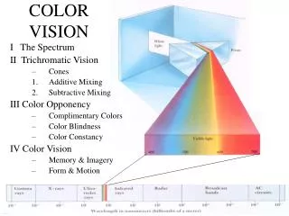
The evolution of color vision. Color vision in Vertebrates. In vertebrates, light is perceived at the level of the retina , a layer of cells at the rear of the eye. The retina contains two types of photoreceptor cells. Cones allow us to distinguish colors.

E N D
The evolution of color vision Jacques van Helden Jacques.van-Helden@univ-amu.fr Aix-Marseille Université (AMU), France Lab. Technological Advances for Genomics and Clinics (TAGC, INSERM Unit U1090) http://tagc.univ-mrs.fr/ FORMER ADDRESS (1999-2011) Université Libre de Bruxelles, Belgique Bioinformatique des Génomes et des Réseaux (BiGRe lab) http://www.bigre.ulb.ac.be/
Color vision in Vertebrates • In vertebrates, light is perceived at the level of the retina, a layer of cells at the rear of the eye. • The retina contains two types of photoreceptor cells. • Cones allow us to distinguish colors. • Rods are not informative about colors, but they are more sensitive than cones and have a higher densit in the retina. They are invoved in the perception of shapes (higher resolution) and night vision (high sensitivity). Illustrations : Jacobs, G. H. and Nathans, J. (2009). The evolution of Primate color vision. Scientific American, 56-63.
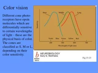
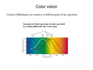
The visible spectrum http://fr.wikipedia.org/wiki/Cône_(biologie) http://en.wikipedia.org/wiki/Visible_spectrum • Each photoreceptor cell perceives a specific range of wavelengths, with a peak at a precise location. • 420 nanometers (nm) for blue-sensitive cones (also called short-wave sensitive: SWS) • 489 nm for the rods • 534 nm for green-sensitive cones (medium-wave sensitive: MWS) • 564 nm for red-sensitive cones (long-wave sensitive: LWS)
Opsins http://www.rcsb.org/pdb/files/1kpn.pdb opsine rétinol Modèle tridimensionnel du pigment des cônes bleus. (Structure PDB 1kpn affichée avec MacPyMol) • The perception of light relies on the presence, in cones and rods, of specialized proteins called opsins. • Opsins bind a small molecule called retinol. The opsin-retinol complex is called rhodopsin. • Rhodopsin is a pigment, which has the capability to capture specific wavelengths in the visible spectra. • The sequence of opsins determines the structure of the protein, which in turn determines the spectrum and the sensitivity of the rhodopsin. • The mutation of a few amino acids at precise positions of the opsin can modify the wavelength that is optimally detected of an opsin (and thus change its spectrum). (Structure PDB 1kpn affichée avec JMol)
Bulls don’t see frogs the same way as we do Illustration: Jacobs, G. H. and Nathans, J. (2009). The evolution of Primate color vision. Scientific American, 56-63. • In Human, color perception relies on 3 types of cones, each having a higher sensitivity to one different color. • Blue (short wavelengths) • Green (medium wavelengths) • Red (long wavelengths) • This kind of color vision relying on 3 photoreceptor types is called trichromatic. • Tichromatic vision can be found in most primates living in Asia and Africa (primates of the « old continent »). • Most other Mammals (with a few exceptions) have a dichromatic vision, relying on two photoreceptors only. • Blue (short wavelengths) • Green (medium wavelengths)
Bulls dont’ see toreros the same way as we do http://ughetto.maisonnave.org/d/42-8/arenes • Here is a simulation of what a bull perceives when it sees the above image. • Traditionally, toreros shake a red cape to stimulate the bulls in bullfighting. • It would make no difference for the bull if the cape would be green instead of red, since rumnants do not distinguish green from red.
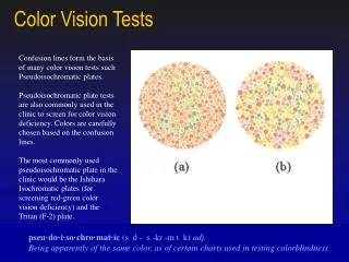
Disruption of some cones provokes daltonism Uneimage colorée Sans les cônes rouges Sans les cônes verts Sans les cônes bleus Vision monochromatique (sans cônes) http://michelf.com/projets/sim-daltonisme/ • Daltonism is a genetic disease that inactivates one or several cone types. • The software tool sim-daltonisme (http://michelf.com/projets/sim-daltonisme/) simulates the effect of daltonism by suppressing some color chanels from a picture.
Relative to birds, all Human beings are Daltonian Pigeon Humain Abeille Etourneau Osorio et al. A review of the evolution of animal colour vision and visual communication signals. Vision Res (2008) vol. 48 (20) pp. 2042-51 • Fishes, reptiles, birds and amphibians have a tetrachromatic vision relying on 4 pigments. • Our capability to discriminate colors is thus weaker than in those species. • We can hardly imagine how a chicken (for example) perceives an image. • Evolution • The ancestor species of all Vertebrates probably had a tetrachromatic vision. • Mammals have lost two types of cones, and their vision thus became dichromatic. • The trichromatic vision of primates results from a secundary acquisition of a third cone (red-sensitive).
Phylogeny of opsins • The phylogenetic tree was buit from the peptidic sequences of a set of opsins. • LWS Long-wave sensitive (red) • SWS Short-wave sensitive (blue/violet/ultra-violet) • Rh1 rods • Rh2 green-sensitive opsin in non-mammalian vertebrates • It allows us to infer the history of the opsin family. Jacobs et al. Evolution of vertebrate colour vision. Clinical & experimental optometry : journal of the Australian Optometrical Association (2004) vol. 87 (4-5) pp. 206-16
Phylogenetic tree of red and blue opsins • We used the Web tool Phylogeny.fr (http://phylogeny.lirmm.fr/), to infer an evolutionary tree from a set of red- and blue-sensitive opsins. Opsine bleue, insectes Mammifères Oiseau Opsine bleue, vertébrés Poissons Un poisson incongru Reptiles Mammifères Oiseau Reptile Bactracien Opsine rouge, vertébrés Lamproies Poissons
Opsin-coding genes • Multi-genomic alignment of the chromosomal region containing LW and MW opsin genes. • Besides the LW gene, we observe two genes in tandem coding for the MW opsin. http://ecrbrowser.dcode.org/xB.php?db=hg18&location=chrX:153138461-153151952