Small Animal Skull
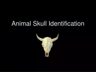
Small Animal Skull. Chapter 15. Skull. Positioning is vital. May have to sedate patient. Remove endotracheal tube Key is precision and symmetry Differences among species and breeds. Anatomy of the Skull. Lateral View.

Small Animal Skull
E N D
Presentation Transcript
Small Animal Skull Chapter 15
Skull • Positioning is vital. • May have to sedate patient. • Remove endotracheal tube • Key is precision and symmetry • Differences among species and breeds
Lateral View • Patient in lateral recumbency with affected side of the skull toward the cassette. • Nasal septum should be parallel to the surface of the cassette. • Mandibular rami should be superimposed. • View should include entire head from the tip of the nose to the base of the skull.
Dorsoventral View • Patient is in sternal recumbency with head resting on cassette. • Front limbs can be in natural position but not in x-ray beam. • May need to put gentle pressure on cervical region or place tape over cranium so that it will remain in desired position. • View should include entire head from tip pf the nose to base of the skull.
Ventrodorsal View • Patient is placed in dorsal recumbency. • Front limbs are extended caudally and secured. • Nose must remain parallel to cassette. • View should include entire head from the tip of the nose to the base of the skull.
Rostrocaudal View of Frontal Sinuses • Patient is in dorsal recumbency with nose pointing upward. • Front legs should be pulled caudally alongside the body. • May need to tie nose in place. • Frontal sinuses should be centered on the cassette. • View should include entire forehead of patient. • Collimator should be aimed perpendicularly to the cassette and centered between the eyes.
Cranium Rostrocaudal view • Dorsal recumbency with nose pointing upward and the front limbs pulled caudally. • Similar to frontal sinus view but nose is directed slightly in a caudal direction (10-15 degrees). • Careful with any endotracheal tube on this view. • Cranium should be centered on the cassette. • View should include entire cranium.
Nasal Cavity- Ventrodorsal Open-mouth view • Patient is in dorsal recumbency with legs extended caudally. • Maxilla remians parallel with the cassette and mouth is held open by tape or gauze. • X-ray tube should be angled 10-15 degrees so that beam is directed into the mouth. • Nasal cavity should be centered on the cassette. • View should include the entire maxilla from the tip of the nose to the pharyngeal region.
Tympanic Bullae-Rostrocaudal Open-mouth view • Patient is placed in dorsal recumbency. • Nose pointing upward and legs pulled caudally alongside the body. • Mouth is tied or propped open. • Nose is pulled cranially 5-10 degrees • View should include the entire nasopharyngeal region of the skull.
Tympanic Bullae-Lateral oblique view • Patient is placed in lateral recumbency • Unaffected bullae toward cassette. • Skull will lay in slightly oblique position.
Temporomandibular Joint-Ventrodorsal Obique View • Patient is in lateral recumbency with affected side toward cassette. • Cranium is rotated approximately 20 degrees toward cassette and held in position. • Can be with mouth opened or closed
Maxilla- Dorsoventral Intraoral View • Patient is placed in sternal recumbency. • Film is placed in mouth • Have to adjust settings as SID has changed. • Now with advent of intraoral radiograph machines, this type of view is less likely on standard machines.
Maxilla- Upper Dental Arcade • Patient is placed halfway on back with maxillary arcade closest to the cassette. • Head is rotated approximately 45 degrees to the cassette. • Mouth should be maintained in open position. • Again new dental machines make this view more rare on standard equipment.
Mandible- Ventrodorsal View • Patient is in dorsal recumbency. • Head is extended in cranial position. • Film is placed in mouth as before. • Adjust SID as before.
Mandible- Lower Dental Arcade • Dorsoventral oblique view. • Patient in lateral recumbency with affected mandible closest to cassette.
Teeth • Will discuss more in depth when we get to dental and intraoral radiographs. • Most accurate way of visualizing teeth. • Patient is in lateral recumbency with unaffected side to the table and affected side up. • Film is inserted into the mouth.