Joints and Body Movement
740 likes | 1.17k Vues
Joints and Body Movement. Introduction to Muscle. Five Golden Rules of Skeletal Muscle Activity. All skeletal muscles cross at least 1 joint. The bulk of a skeletal muscle lies proximal to the joint crossed. All skeletal muscles have at least 2 attachments: origin and insertion.

Joints and Body Movement
E N D
Presentation Transcript
Joints and Body Movement Introduction to Muscle
Five Golden Rules of Skeletal Muscle Activity • All skeletal muscles cross at least 1 joint. • The bulk of a skeletal muscle lies proximal to the joint crossed. • All skeletal muscles have at least 2 attachments: origin and insertion. • Skeletal muscles can only pull they never push. • During contraction, a skeletal muscle insertion moves toward the origin.
Muscles and Body Movements • Movement is attained due to a muscle moving an attached bone • Muscles are attached to at least two points • Origin • Attachment to a moveable bone • Insertion • Attachment to an immovable bone
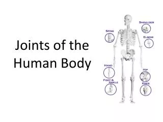
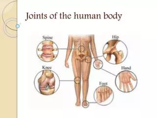
Joints • Definition of joint • Area where two bone articulate (come together) • Two major functions • Hold bones together • Allow for mobility – fewer joints produce robot type motion. • Classification • Functionally – degree of motion allowed • Structurally - based on tissue and anatomy of the joint
Functional Classification • Synarthrosis • no movement (sutures, syndesmosis, gomphosis) • Amphiarthrotic • Slight degree of movement (sychondrosis, symphsis) • Diarthrotic • Freely moveable • Differ from one another in terms of specific degrees of movement allowed between bony surfaces • Six types – Hinge, Pivot, Condyloid, Saddle, Ball and socket, Gliding or plane.
Structural Classification - Fibrous • Bones held together by dense collagen fibers with little elasticity and no spaces between bones • 3 types • Sutures – irregular edges of bone held together by short fibers, not moveable, skull bones • Syndesmoses – bones connected by a long fibrous connective tissue which allows for a slight amount of movement – interosseous membrane in arm and leg • Gomphosis – tooth attachment to maxilla or mandible – specialized ligaments are strong and cause joint to be immoveable
Structural - cartilaginous • Tissue made of collagen which has a gel-like quality making it flexible and strong. A great shock absorber. • 2 kinds • synchondrosism • growth plate • between first rib and sternum • between manubrium and sternal body • Symphyses • greater elasticity and flexibility • Found between vertebrae – allows movement but keeps bones in place. • Also pubic area – pubic symphasis
Structural - synovial • Articulating bone ends are separated by a joint cavity which contains synovial fluid • Synovial capsule is lined with a smooth connective tissue membrane. • Articulating surfaces are covered with hyaline cartilage • Articular capsule is reinforced with ligaments • Bursa sacs of the tendon sheaths reduce friction where the ligament and muscle cross the bone. • All are freely moveable in single or multiple planes
Types of synovial joints • Hinge • Concave surface on a convex surface • Elbow , knee • Uniaxial movement – usually flexion and extension • Pivot • One bone rotates on a fixed landmark • Atlas and axis, radius and ulna • Uniaxial rotation • Condyloid • Oval condyle of one bone fits into an oval depression in another • Mandible on temporal bone, metacarpal on proximal phalange • Biaxal –two way movement
Types of Synovial Joints • Saddle • Articulating bones are saddle shaped • Joint between the thumb and trapezium (carpal) bone • Movement is biaxial – two planes • Gliding or plane • 2 semi flat surfaces facing one another • Facets of the vertebrae: intercarpal and intertarsal joints • Sliding movements • Ball and Socket • Round sphere fits into a cup • Shoulder, hip • Movement in all directions
Inflammatory Conditions Associated with Joints • Bursitis – inflammation of a bursa; usually caused by a blow or friction • Tendonitis – inflammation of the tendon sheaths due to overuse • Arthritis – inflammatory or degenerative diseases of joints • Osteoarthritis – most common chronic form due to normal aging • Rheumatoid – autoimmune disease – Immune system attacks joints • Gouty arthritis – inflammation of joints is caused by uric acid deposits crystallized from the blood – treated with dietary changes.
Types of Body Movements • Flexion • Angle between 2 body parts decreases • Brings two bones closer together • Typical of hinge joints like knee and elbow • Extension • Angle between 2 body parts increases • Hyperextension • Excessive increase in angle between 2 body parts • Lateral flexion • Bending the vertebral column to the right or left of midline
Body Movements • Rotation • Movement of a bone around its longitudinal axis • Common in ball-and-socket joints • Example is when you move atlas around the dens of axis (shake your head “no”)
Body Movements • Abduction • Movement of a limb away from the midline • Adduction • Opposite of abduction • Movement of a limb toward the midline • Circumduction • Combination of flexion, extension, abduction, and adduction • Extremity moves in a large circle • Common in ball-and-socket joints
Body Movements • Dorsiflexion • Lifting the foot so that the superior surface approaches the shin – pointed skyward • Plantar flexion • Extend ankle until toes downward – toes to floor
Body Movements • Inversion • Turn sole of foot medially - inward • Eversion • Turn sole of foot laterally – to the side
Body Movements • Supination • Forearm rotates laterally so palm faces up • Pronation • Forearm rotates medially so palm faces downward
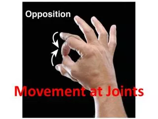
Body Movements • Opposition • Move thumb to touch the tips of other fingers on the same hand
Body Movements • Elevation • to move a body part up like shrugging shoulders. • Depression • to move body part downward – pushing shoulders down • Protraction • move body part forward like your jaw • Retraction • move body part to the back like your jaw
Types of Muscles • Prime mover—muscle with the major responsibility for a certain movement • Antagonist—muscle that opposes or reverses a prime mover • Synergist—muscle that aids a prime mover in a movement and helps prevent rotation • Fixator—stabilizes the origin of a prime mover
Naming Skeletal Muscles • By direction of muscle fibers • Example: Rectus (straight) • By relative size of the muscle • Example: Maximus (largest) • By location of the muscle • Example: Temporalis (temporal bone) • By number of origins • Example: Triceps (three heads) • By action of the muscle • Example: flexor or extensor – flexes or extends a bone
Naming Skeletal Muscles • By location of the muscle’s origin and insertion • Example: Sterno (on the sternum) • By shape of the muscle • Example: Deltoid (triangular)
Head and Neck Muscles • Frontalis • raises eyebrows • Origin – cranial aponeurosis • Insertion – Skin of eyebrows and nose • Occipitalis • Pulls scalp posteriorly • Origin – occipital and temporal bone • Insertion – cranial aponeurosis • Orbicularis oculi • closes eyes, squints, blinks, winks • Origin – Frontal and maxillary bone • Insertion – tissue of eyelids
Head and Neck Muscles • Orbicularis oris • Action -closes mouth and protrudes the lips • Origin – Maxilla and Mandible • Insertion – Muscle and skin at angle of mouth • Buccinator • Action - flattens the cheek, chews • Origin – maxilla and mandible • Insertion – obicularisoris • Zygomaticus • Action - raises corners of the mouth • Origin – Zygomatic bone • Insertion – Skin and muscle at corner of mouth
Head and Neck Muscles • Masseter • Action - closes the jaw and elevates mandible • Origin – Zygomatic process • Insertion – Mandible • Temporalis • Action - synergist of the masseter, closes jaw • Origin – Temporal bone • Insertion - Mandible • Platysma • Action -pulls the corners of the mouth inferiorly • Origin – Fascia of chest • Insertion – Lower edge of mandible
Head and Neck Muscles • Sternocleidomastoid • Action - flexes the neck, rotates the head • Origin – Sternum and Clavicle • Insertion – Mastoid process • Sternohyoid • Action – depresses larynx and hyoid bone • Origin - manubrium • Insertion – hyoid bone
Muscles of the Shoulder • Trapezius • Action – Extends neck, adducts scapula • Origin – Occipital bone, cervical and thoracic vertbrae • Insertion – Acromion and spinous process of scapula, clavicle • Deltoid • Action – arm abduction, flexion, extension and rotation of humerus • Origin – clavicle, acromion, spine of scapula • Insertion – Deltoid tuberosity of humerus
Muscles of the Shoulder • Infraspinatus • Action – rotates humerus laterally • Origin – scapula • Insertion – greater tubercule of humerus • Teres minor • Action – rotates humerus laterally • Origin – scapula • Insertion – greater tubercule of humerus • Teres major • Action – Extends, rotates and adducts humerus • Origin – scapula • Insertion – lesser tubercle
Muscles of Arm • Triceps brachii • Action – extends lower arm • Origin – glenoid cavity, posterior humerus • Insertion – olecranon process of ulna • Biceps brachii • Action –flexes elbow and supinates forearm • Origin – coracoid process of scapula • Insertion – Proximal radius • Brachialis • Action – major arm flexion • Origin – anterior surface of distal humerus • Insertion – Coronoid process of ulna
Muscles of Forearm • Pronator teres • Action – pronates forearm • Origin – Distal humerus and coronoid process of ulna • Insertion – Radius • Brachioradialis • Action – Forearm flexion • Origin – Distal humerus • Insertion – Styloid process of radius • Flexor carpi radialis • Action – flexes wrist and abducts hand • Origin – medial epicondyle of humerus • Insertion – Second and third metacarpals
Muscles of Forearm • Palmaris longus • Action – weak wrist flexor • Origin – medial epicondyle of humerus • Insertion – fascia of palm • Extensor carpi radialis longus • Action – extends wrist, abducts wrist • Origin – lateral condylar ridge of humerus • Insertion – second metacarpal • Flexor carpi ulnaris • Action – flexes wrist, adducts hand • Origin – Distal humerus and posterior ulna • Insertion – Fifth metacarpal and carpals
Muscles of Forearm • Extensor digitorium • Action – extends finger, extends wrist • Origin – Lateral epicondyle of humerus • Insertion – Distal phalanges of 2-5 finger • Extensor carpi ulnaris • Action – Extends and adducts wrist • Origin – Lateral epicondyle of humerus • Insertion – Fifth metacarpal
Thorax Muscles • Pectoralis major • Action – flexes arm, adducts and medially rotates arm • Origin – clavicle, sternum, and cartilare of first 6 ribs • Insertion – greater tubercle of humerus • Serratus anterior • Action – Rotates scapula • Origin – ribs 1 – 8 • Insertion – anterior surface of medial border of scapula
Abdominal muscles • Rectus abdominus • Action – Flex and rotate lumbar region of vertebrae • Origin – pubic crest and pubic symphysis • Insertion – Xiphoid process and costal cartilage of ribs 5-7 • External oblique • Action – flex vertebral column and compress abdominal wall, trunk rotation and lateral flexion • Origin – lower eight ribs • Insertion – linea alba, pubic crest, iliac crest
Abdominal muscles • Internal oblique • Action - flex vertebral column and compress abdominal • Origin – lumbar fascia • Insertion – linea alba, pubic crest, last 3 ribs • Transverse abdominus • Action – compresses abdominal contents • Origin – inguinal ligament, lumbar fascia, cartilages of last 6 ribs, iliac crest • Insertion – linea alba, pubic crest
Hip Muscles • Gluteus medius • Action – Abducts, and medially rotates thigh; steadies pelvis while walking • Origin – side of illium • Insertion – greater trochanter of femur • Gluteus maximus • Action – hip extender – climbing • Origin – Ilium, sacrum and coccyx • Insertion – Gluteal tuberosity of femur