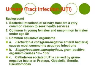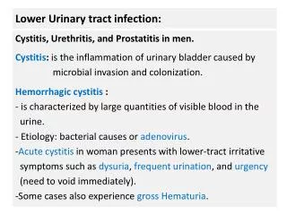
Lower Urinary tract infection:
Lower Urinary tract infection:. Cystitis, Urethritis, and Prostatitis in men. Cystitis: is the inflammation of urinary bladder ( the sac that temporarily holds urine until urination) caused by microbial colonization and infection.

Lower Urinary tract infection:
E N D
Presentation Transcript
Lower Urinary tract infection: Cystitis, Urethritis, and Prostatitis in men. Cystitis: is the inflammation of urinary bladder (the sac that temporarily holds urine until urination) caused by microbial colonization and infection. Hemorrhagic cystitis : is characterized by large quantities of visible blood in the urine. It can be caused by an infection (bacterial or adenovirus). Urethritis : It is the inflammation of urethra ( the tube that carries urine from the bladder during urination) caused by microbial infection.
n Symptoms include a burning sensation during urination and fever. Most of the cases of urethritis without cystitis are sexually transmitted.
a Chronic bacterial prostatitis: The prostate is a small gland and part of the male reproductive system. It produces fluid that becomes part of a man's semen, the liquid that helps transport sperm. If the prostate becomes inflamed, the condition is called prostatitis.
Etiology of Cystitis and Urethritis (Non-Sexual origin): Causes: Pathogens may ascending the urethra of fecal origin. 1- Escherichia coli. 2- Staphylococcus saprophyticus. 3- Other genera of Enterobacteriaceae : Klebsiella, Enterobacter, Proteus, and Serratia. 4- Pseudomonas aeruginosa ( Hospital-acquired infection due to contaminated catheter). 5- Enterococcus faecalis . ( Hospital-acquired infection due to contaminated catheter).
Laboratory diagnosis of UTI: Collection of Clinical Specimens: Urine specimens: A- Midstream catch urine: One to Ten ml: must be collected in sterile urine container , the first portion must be discarded to avoid contamination with urethra and vaginal bacteria. B- Catheterized Urine: One to Ten ml Must be collected in sterile urine container. C- Suprapubic Urine: One to Ten mL Aspirated from urinary bladder by fine needle collected in sterile anaerobic tube. ( young babies, children, obstruction cases).
Cultivation of urine specimens: Urine specimens should be cultivated on Enriched media (blood agar) and differential media (MacConkey’s agar or CLED) by the following methods: 1- Four quadrant streaking method: depends on Calibrated Loop. :( Semi-quantitative). 2- Network Streaking method: depends on Calibrated Loop. : ( quantitative). 3- Microbroth dilution method. ( quantitative) Calibrated loop carry only one microliter.
Isolation of microbe from urine and calculation of CFU : Bacterial loop carry one microlitre. When 50 colonies are isolated = 50cellsper/1 µl = 50 CFU/ µl In onemL: 50 * 1000= 50000 CFU/mL.
CFU cutoff value and significant bacteriuria: The CFU cutoff value is associated with type of urine specimens. It is calculated to be equal to: For Midstream catch urine:50000- 100000 CFU/m L. For Catheterized Urine: 100 CFU/m L. For Suprapubic Urine: Any growth. Significant bacteriuria: Is the isolation of microbes of infectious dose cutoff value from urine specimen in presence of signs and symptoms of UTI. It is mainly associated with pyuria and dysuria.
Isolation of Enterobacteriaceaefrom urine specimens: The most important genera that could be isolated from urine are: Escherichia coli ,Klebsiella, Enterobacter, Proteus mirabilis, and Serratia species. Enterobacteriaceaegenera are classified according to lactose fermentation into two groups: 1-Lactose non-fermenters. 2- Lactose fermenters.
Identification of E. coli: -Some strains of uropathogenic E. coliare verotoxin producers. -All strains are oxidase negative and catalase positive. -Metallic green sheen on EMB agar. -Uropathogenic E. coli species are indole positive.
Identification of Proteus species: All species are Gram’s negative bacilli that illustrate swarming growth on blood agar. All are H2S and urease positive.
Isolation of Pseudomonasspecies from urine specimens: Cultural characteristics: -All species of Pseudomonas produce Water soluble Exopigmenton nutrient agar.(greenish yellowish pyoverdin) . -All species are oxidase positive.
Isolation of Serratia marcescens from urine: Microorganisms that produce pigment during growth are said to be chromogenic. Serratia marcescens produces a deep red pigment ( water-insoluble) at 25°C, but does not produce pigment at 37°C. Serratia marcescens Pseudomonas
Isolation of Gram’s positive Cocci : -The most important Gram’s positive Cocci that associated with UTI are: 1-Staphylococcussaprophyticus. 2-Enterococcusfaecalis. -Staphylococcus aureus isalso considered as a causative agent of UTI. Differentiation of Staphylococcus aureusand other Staphylococcus species: -All Staphylococcus species are Gram’s positive Cocci arranged in clusters. -All species are Catalase positive. -Staphylococcus aureusare coagulase positive. -Staphylococcus epidermidisand Staphylococcus saprophyticusare coagulase negative.
Staph. aureus coagulase positive Other Staph. Coagulase negative.
n -Staphylococcus saprophyticusspecies are Mannitolfermenters and Novobiocinresistant. Novobiocin sensitive Staph. Mannitol fermentation pattern.
Isolation of Enterococcus faecalisfrom urine specimens: Urine specimens should be cultivated on bile esculine salt agar when Nosocomial UTI is suspected. Black discoloration of media is a distinctive characteristics of group D streptococcus growth ( Enterococcus faecalis).
Parasitic infection of urinary system: Schistosomiasis: Classification (phylum): Trematoda. Classification according to morphology: Flattened non-segmented unisexual flukes. Schistosoma hematobium: • Found in Middle East, Africa, Portugal and Cyprus • Definitive host: man • Intermediate host: snail • It causes urinary bilharziasis (female deposits eggs in the vesical and pelvic plexus of venous circulation).
n Male: 10-20 x I mm with short anterior cylindrical pan and posterior flattened part which incurved ventrally to form the gynaeccphoric canal. It has well developed oral and ventral suckers. Female: 15-25 x 0.25 mm:, cylindrical, ovary may "be central, anterior or posterior, uterus may be short or long containing eggs. Schistosoma hematobiumegg: -140x50 µ, oval with terminal spine. -Passes in urine.
CLINICAL PICTURE: The clinical picture is divided into three stage: 1-Incubation period; (acute Schistosomiasis 6-12 Weeks) : This stage is characterized by skin irritation after coming from water, and coughing. Late in the incubation period, patients complains of enlarged liver, fever, headache, anorexia, loss of weight. CBC shows Eosinophilia. 2- Stage of egg deposition: (1- 5 years after infection). Urinary bilharziasis: caused by S.hematobium,characterized by terminal haematuria, burning sensation during urination.
n 3-Stage of complication: -Due to a fibrosis around the eggs in bladder, liver and lung. - Obstruction of bladder. Urinary bilharziasis: cystitis, hydronephrosis and Pyonephrosis, sterility in males, renal failure, urinary bladder carcinoma, pulmonary hypertension and heart failure.