Download
1 / 12
120 likes | 264 Vues
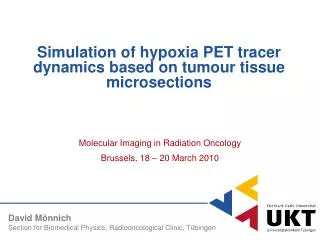
Simulation of hypoxia PET tracer dynamics based on tumour tissue microsections. Molecular Imaging in Radiation Oncology Brussels, 18 – 20 March 2010. David Mönnich Section for Biomedical Physics, Radiooncological Clinic, Tübingen. Introduction. Larynx carcinoma, FMISO PET 4 h p.i.

E N D
Simulation of hypoxia PET tracer dynamics based on tumour tissue microsections Molecular Imaging in Radiation Oncology Brussels, 18 – 20 March 2010 David Mönnich Section for Biomedical Physics, Radiooncological Clinic, Tübingen
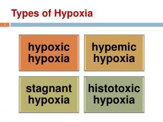
Introduction Larynx carcinoma, FMISO PET 4 h p.i. • Dose distribution can be tailored by IMRT (Dose Painting) • Hypoxia PET imaging with [18F]-FMISO • Dynamic PET imaging: Voxel-wise Time Activity Curves (TACs) TACs Perfusion peaks D. Mönnich, MIRO 2010, Brussels 2
Objective • Link PET voxel signals to the underlying tumour microstructure ? ? D. Mönnich, MIRO 2010, Brussels 3
Creating vascular maps from tumour microsections • Extractedvascularmap • Regionswith different vascularfractions (VF) 3.9 mm Low VF (1.0%) Interm. VF (3.4%) High VF (14.5%) Courtesy of E. Troost, UMC Nijmegen Blue: Endothelium Red: Proliferation Green: Hypoxia D. Mönnich, MIRO 2010, Brussels 4
Model & Simulation Method Solve oxygen reaction-diffusion equation Static oxygen distribution D. Mönnich, MIRO 2010, Brussels 5
Model & Simulation Method Solve oxygen reaction-diffusion equation Necrotic regime Increase Static oxygen distribution D. Mönnich, MIRO 2010, Brussels 6
Model & Simulation Method Solve oxygen reaction-diffusion equation Solve FMISO reaction-diffusion equation Necrotic regime Increase Static oxygen distribution D. Mönnich, MIRO 2010, Brussels 7
Results • Tracer supply to hypoxic areas is diffusion limited • FMISO binding is not hypoxia specific in the first 15 minutes 3.8 mm D. Mönnich, MIRO 2010, Brussels 8
Results • Tracer supply to hypoxic areas is diffusion limited • FMISO binding is not hypoxia specific in the first 15 minutes 3.8 mm D. Mönnich, MIRO 2010, Brussels 9
Results • TACs from differently perfused regions D. Mönnich, MIRO 2010, Brussels 10
Results • TACs from differently perfused regions • Different microstructure can lead to similar late signal D. Mönnich, MIRO 2010, Brussels 11
Summary and conclusion • Summary • FMISO supply to hypoxic regions is limited by molecular diffusion speed on relevant time scales • Different tissue microstructure can lead to similar voxel activitiy in static PET images • Conclusion • Dose painting using static images might omit hazardous tumour regions • Imaging of perfusion seems necessary in addition to static hypoxia imaging D. Mönnich, MIRO 2010, Brussels 12


![PET Tracer Coordination [ F-18] labeled](https://cdn3.slideserve.com/6693342/slide1-dt.jpg)