Download
1 / 41
430 likes | 669 Vues
Neurologic and Musculoskeletal Imaging Studies Pediatric MSK radiology2. دکترامیر هوشنگ واحدی متخصص طب فیزیکی و توانبخشی قسمت 6. AVN. (Osteonecrosis Or aseptic necrosis).

E N D
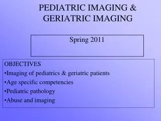
Neurologic and Musculoskeletal Imaging StudiesPediatric MSK radiology2 دکترامیر هوشنگ واحدی متخصص طب فیزیکی و توانبخشی قسمت 6
AVN (Osteonecrosis Or aseptic necrosis)
Legg-Perthes disease.A, fragmentation and sclerosis of the right femoral epiphysis in this 6-year-old boy. B, A follow-up film obtained 8 years later shows continuing deformity due to the osteonecrosis. Significant degenerative arthritis (C) developed by age 12 years.
Avascular Necrosis (AVN) of the Hip. Subchondral lucency (arrows) is seen in the weight-bearing portion of this hip with AVN. Patchy sclerosis throughout the femoral head is also noted.
Geode in the Hip. A large cystic lesion (arrows) is seen in this patient with avascular necrosis (AVN) of the hip. Note the adjacent patchy sclerosis indicative of AVN. A subchondral cyst or geode should be considered any time a lytic lesion is found around a joint
AVN of the Shoulder. Articular surface collapse is present in this shoulder with long-standing AVN
Sever’s Disease, calcaneal apophysitis, is a common and painful condition experienced by growing children.
Scheuermann Disease. Avascular necrosis of the apophyseal rings of the vertebral bodies is called Scheuermann disease. He originally described a painful kyphosis with multiple vertebral bodies involved.
Mild wedge compression deformities of multiple thoracic vertebrae are present, resulting in mild kyphosis. (B) disk spaces are narrowed. Vertebral end-plate contour is undulating with radiolucent depressions and adjacent sclerosis due to intraosseous prolapse of disc material, which is commonly referred to as Schmorl's nodes (arrows).
Freiberg Infraction. Flattening, collapse, and sclerosis of the second metatarsal head,as seen in this patient, is typical of avascular necrosis or Freiberg infraction. It can also involve the second, third, or fourth metatarsal heads.
Köhler Disease. Flattening and sclerosis of the tarsal navicular (arrow) in children is thought by many to be avascular necrosis and is called Köhler disease
Kienböck Malacia. The increased density and partial fragmentation of the lunate are characteristic for AVN
Osteochondritis Dissecans. A small focal area of (AVN) in the medial epicondyle of the femur (black arrows) is present, which is an area of osteochondritis dissecans. Part of the area of AVN has shed a bony fragment (white arrow) that is loose in the joint, which is known as a loose body or “joint mouse.”
Osteochondritis Dissecans of the Talus. A focal area of avascular necrosis in the talus, as seen here (arrows), is called osteochondritis dissecans. The talus is the second most common site after the knee and, as in the knee, can cause a joint mouse, or loose body in the joint.
Osteochondritis Dissecans of the Elbow. The third most common site for osteochondritis dissecans is in the capitellum of the elbow. The faint lucency seen in this capitellum (arrows)
Slipped Capital Femoral Epiphysis SCFE Klein's Line • Line drawn along superior border of femoral neck should cross at least a portion of the femoral epiphysis • Most sensitive indicator of a mild slip on plain film Classification Grade I: displacement of epiphysis less than 30% of width of femoral neck Grade II: slip between 30%-60% Grade III: includes slips of greater than 60% the width of neck
When Klein’s Line Fails, Try Capener’s Sign • On PA, ischium and femoral head overlap to yield crescent of double density • SCFE reduces overlap area • Sometimes more sensitive than Klein’s line alone