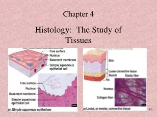
Tissues: The living fabric
630 likes | 825 Vues
Tissues: The living fabric. Ch 4 a Epithelial Tissue. Tissues. Histology = Study of tissues Tissue = Groups of cells similar in structure and function The four types of tissues Epithelial - covering Connective - support Muscle - movement Nerve - control. Epithelial Tissue. 2 types

Tissues: The living fabric
E N D
Presentation Transcript
Tissues: The living fabric Ch 4 a Epithelial Tissue
Tissues • Histology = Study of tissues • Tissue = Groups of cells similar in structure and function • The four types of tissues • Epithelial - covering • Connective - support • Muscle - movement • Nerve - control
Epithelial Tissue • 2 types • Covering and lining epithelium • Glandular epithelium
Epithelial Tissue • A sheet of cells that covers a body surface or lines a body cavity • Forms boundaries between different environments
Epithelial Tissue Functions • Protection • Absorption • Filtration • Excretion • Secretion (from glands) • Sensory reception
Characteristics of Epithelial Tissue • Cellularity – composed almost entirely of cells • Very little extracellular material • Special contacts – form continuous sheets held together by tight junctions and desmosomes
Characteristics of Epithelial Tissue • Polarity – apical and basal surfaces • Apical = free upper surface exposed to exterior of body or cavity (some are slick, some have cilia, most have microvilli) • Basal = lower surface attached
Characteristics of Epithelial Tissue • Basal surface attached to basement membrane • basal laminae – thin, adhesive, non-cellular • reticular laminae – fine network of collagen fibers belonging to the connective tissue underneath, non-cellular
Characteristics of Epithelial Tissue • All epithelial tissue are supported by and rest upon connective tissue
Characteristics of Epithelial Tissue • Avascular but innervated – contains no blood vessels but supplied by nerve fibers – nourished by diffusion • Regenerative – rapidly replaces lost cells by cell division
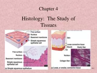
Classification of Epithelia • Simple • stratified Figure 4.1a
Classification of Epithelia • Squamous • Cuboidal • Columnar Figure 4.1b
Epithelia: Simple Squamous • Single layer of flattened cells with disc-shaped nuclei and sparse cytoplasm
Epithelia: Simple Squamous • Functions • Diffusion and filtration • Provide a slick, friction-reducing lining in lymphatic and cardiovascular systems • Present in the kidneys, lungs, lining of heart, blood vessels, lymphatic vessels
Epithelia: Simple Squamous Figure 4.2a
Epithelia: Simple Cuboidal • Single layer of cubelike cells with large, spherical central nuclei • Function in secretion and absorption • Present in kidney tubules, ducts and secretory portions of small glands, and ovary surface
Epithelia: Simple Cuboidal • Single layer of cubelike cells with large, spherical central nuclei • Function in secretion and absorption • Present in kidney tubules, ducts and secretory portions of small glands, and ovary surface Figure 4.2b
Epithelia: Simple Columnar • Single layer of tall cells with oval nuclei; many contain cilia • Goblet cells are often found in this layer • Cells that secrete a protective lubricating mucus • Function in absorption and secretion
Epithelia: Simple Columnar • Nonciliated type line digestive tract and gallbladder • Ciliated type line small bronchi, uterine tubes, and some regions of the uterus • Cilia help move substances through internal passageways
Epithelia: Simple Columnar Figure 4.2c
Epithelia: Pseudostratified Columnar • Single layer of cells with different heights; some do not reach the free surface • Nuclei are seen at different layers
Epithelia: Pseudostratified Columnar • Function in secretion and propulsion of mucus • Present in the male sperm-carrying ducts (nonciliated) and trachea (ciliated)
Epithelia: Pseudostratified Columnar • Single layer of cells with different heights; some do not reach the free surface • Nuclei are seen at different layers • Function in secretion and propulsion of mucus • Present in the male sperm-carrying ducts (nonciliated) and trachea (ciliated) Figure 4.2d
Epithelia: Stratified Squamous • Thick membrane composed of several layers of cells • Function in protection of underlying areas subjected to abrasion
Epithelia: Stratified Squamous • Forms the external part of the skin’s epidermis (keratinized cells), and linings of the esophagus, mouth, and vagina (nonkeratinized cells)
Epithelia: Stratified Squamous • Thick membrane composed of several layers of cells • Function in protection of underlying areas subjected to abrasion • Forms the external part of the skin’s epidermis (keratinized cells), and linings of the esophagus, mouth, and vagina (nonkeratinized cells) Figure 4.2e
Epithelia: Stratified Cuboidal • Stratified cuboidal • Quite rare in the body • Found in some sweat and mammary glands • Typically two cell layers thick
Epithelia: Stratified Columnar • Stratified columnar • Limited distribution in the body • Found in the pharynx, male urethra, and lining some glandular ducts • Also occurs at transition areas between two other types of epithelia
Epithelia: Transitional • Several cell layers, basal cells are cuboidal, surface cells are dome shaped • Stretches to permit the distension of the urinary bladder • Lines the urinary bladder, ureters, and part of the urethra
Epithelia: Transitional • Several cell layers, basal cells are cuboidal, surface cells are dome shaped • Stretches to permit the distension of the urinary bladder • Lines the urinary bladder, ureters, and part of the urethra Figure 4.2f
Epithelia: Glandular • A gland is one or more cells that makes and secretes an aqueous fluid
Epithelia: Glandular • Glands Classified by: • Site of product release – • Endocrine (internally secreting) or Exocrine (externally secreting) • Relative number of cells forming the gland – • Unicellular (one cell) & Multicellular (many cells)
Endocrine Glands • Ductless glands that produce hormones • Secretions include amino acids, proteins, glycoproteins, and steroids
Exocrine Glands • More numerous than endocrine glands • Secrete their products onto body surfaces (skin) or into body cavities
Exocrine Glands • Examples include mucous, sweat, oil, and salivary glands, the liver (which secrets bile), the pancreas (which secrets enzymes) and others
Unicellular Exocrine Glands • The only important unicellular exocrine gland is the goblet cell • Found in linings of intestinal and respiratory tracts • Secrets mucus
Multicellular Exocrine Glands • Multicellular exocrine glands are composed of a duct and secretory unit • Classified according to: • Simple or compound duct type • Structure of their secretory units
Structural Classification of Multicellular Exocrine Glands Figure 4.3a-d
Structural Classification of Multicellular Exocrine Glands Figure 4.3e-g
Modes of Secretion • Merocrine – products are secreted by exocytosis (e.g., pancreas, sweat, and salivary glands) • Holocrine – products are secreted by the rupture of gland cells (e.g., sebaceous glands)
Modes of Secretion Merocrine Holocrine Figure 4.4