Download
1 / 21
360 likes | 1.21k Vues
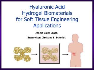
D irecting Tissue Regeneration via Hyaluronic Acid Hydrogel Scaffolds. Jennie Baier Leach 1 , Charles W. Patrick, Jr. 2,3 , Christine E. Schmidt 2. 1 Department of Chemical Engineering 2 Department of Biomedical Engineering The University of Texas at Austin, Austin, TX

E N D
Directing Tissue Regeneration via Hyaluronic Acid Hydrogel Scaffolds Jennie Baier Leach1, Charles W. Patrick, Jr.2,3, Christine E. Schmidt2 1 Department of Chemical Engineering 2 Department of Biomedical Engineering The University of Texas at Austin, Austin, TX 3 Department of Plastic Surgery The University of Texas M. D. Anderson Cancer Center, Houston, TX
Overall goal:To create an angiogenic hydrogelthat is specifically designed to promote tissue repair Figures from http://www-2.cs.cmu.edu/People/tissue/tutorial.html
“Biomaterials that heal” Ratner BD. (2002) J Controlled Release. 78:211-8 Aim: To facilitate natural wound healing biology Biological design: Biomimetic molecules like those present in a wound Limited nonspecific protein adsorption Nonimmunogenic Materials design: Enzymatic degradation Versatile modification strategies www.organogenesis.com Hubbell JA. (1999) Curr Opin Biotech. 10:123-9; Stocum DL. (1998) Wound Repair Regen. 6:276-90
Nerve Regeneration Nerve cable Blood vessels Mackinnon SE & Dellon AL, Surgery of the Peripheral Nerve, Thieme Medical (1988) Adapted from http://www.ama-assn.org/ama/pub/category/7172.html
Specific goal: To develop and characterize hyaluronic acid (HA) hydrogel scaffolds Hyaluronic acid
Biomaterials: State of the Art Proteins Fibrin, Collagen “Sugars” Agarose, Alginate Chitosan, Dextran, HA Synthetic polymers Polyethylene glycol (PEG) Polylactic-co-glycolic acid (PLGA) Polyhydroxyethyl methacrylate (pHEMA) Inherent biological activity Nonimmunogenic Multiple modification sites Tunable material properties
HA’s role in wound healing Chen WY & Abatangelo G. (1999) Wound Repair Regen. 7:79-89.
Hyaluronic Acid (HA) glucuronic acid acetylglucosamine • Natural ECM component • Inherently angiogenic upon degradation • Non-immunogenic • Multiple sites available for modification
Cross-linked HA hyase hyase • Combines controlled delivery and physical support scaffold • Slows down natural degradation by hyaluronidase • - Maintains HA’s hydrated, porous structure Cross-linked HA Hydrogels Native HA hyase hyase
glycidyl methacrylate (GM) + Photoinitiator + UV light Cross-linked GMHA GMHA HA GM modification HA Methacrylation and Cross-linking Baier Leach J, Bivens KA, Patrick CW, Jr. & Schmidt CE. (in revision) Biotech Bioeng. Cross-linking Variables: GMHA concentration (0.5-2.0%) UV exposure (1-4 min, ~22 mW/cm2) Photoinitiator conc. (0.03-3% Irgacure 2959) % methacrylation (NMR: ~5-11%)
5% 7% 11% Increasing % methacrylation Enzymatic Degradation Rate GMHA Solution +UV Incubate in hyase Measure weight loss of gel over time Remove mold
Media Hydrogel Crosslinked GMHA 1% GMHA 0.1% Irgacure 2959 0.03% N-vinyl pyrrolidinone 1 minute UV Crosslinked GMHA GMHA in solution Media HA HAEC Cytocompatibility Method adapted from Trudel J & Massia SP. (2002) Biomaterials. 23:3299-3307. HAEC monolayer www.corning.com
3. Harvest tissue at 2 weeks (side view without TEC) 1. Fill TEC with hydrogel 4 hydrogels per rat + Control: Fibrin -Control: Agarose 2 HA gels 2. Suture to muscle 4. EC immunostain (CD31) In Vivo Analysis of Angiogenicity Method from King TW, Brey EM, Youssef AA, Johnston C & Patrick CW, Jr. (2002) Anal Quant Cytol Histol. 24:39-48.
In Vivo Analysis of Angiogenicity Fibrin (+) GMHA hydrogel 6.63 ± 1.10, n = 9 (% area CD31-positive cells)7.06 ± 0.14, n = 4 2 Week Implant Scale, 200 mm Agarose (-)
HAEC adhesion on GMHA-RGD RGD bound to GMHA hydrogel using EDC/NHS (Method adapted from alginate studies in Rowley JA, Madlambayan G & Mooney DJ. (1999) Biomaterials. 20:45-53.) GMHA, no RGD GMHA-RGD 0.23 mol RGD/mol GMHA disaccharide or 0.61 mM RGD in hydrogel 250 mm
Summary GMHA hydrogels are photopolymerizable, degradable, cytocompatible, angiogenic (comparable to fibrin in subcutaneous implants) and can be modified with RGD. • Future Work • Tune angiogenicity of HA hydrogels: • Vary adhesive peptides concentrations (KGHK, REDV) • Encapsulate growth factors (VEGF, bFGF)
Specific goal: To develop and characterize hyaluronic acid (HA) hydrogel scaffolds Hyaluronic acid
Overall goal:To create an angiogenic hydrogelthat is specifically designed to promote tissue repair Figures from http://www-2.cs.cmu.edu/People/tissue/tutorial.html
Nerve Regeneration Nerve cable Blood vessels Mackinnon SE & Dellon AL, Surgery of the Peripheral Nerve, Thieme Medical (1988) Adapted from http://www.ama-assn.org/ama/pub/category/7172.html
AcknowledgementsClear Solutions Biotech Stoney Brook, NY Dr. C.P. Pathak Sulzer Biologics, Austin TXCarol Johnston The U.T.M.D. Anderson Cancer Center, Houston, TXFunding AgenciesNIH Biotechnology Training Grant Fellowship (JBL)NIH HL62341 (CWP), CA1667 (UTMDACC)The Whitaker Foundation (CWP)NSF Grant BES-9733156 (CES) Gillson-Longenbaugh Foundation Grant (CES)