Download
1 / 50
510 likes | 1.19k Vues
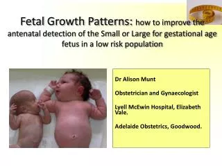
Fetal Growth Patterns: how to improve the antenatal detection of the Small or Large for gestational age fetus in a low risk population. Dr Alison Munt Obstetrician and Gynaecologist Lyell McEwin Hospital, Elizabeth Vale. Adelaide Obstetrics, Goodwood. Fetal Growth Patterns.

E N D
Fetal Growth Patterns: how to improve the antenatal detection of the Small or Large for gestational age fetus in a low risk population Dr Alison Munt Obstetrician and Gynaecologist Lyell McEwin Hospital, Elizabeth Vale. Adelaide Obstetrics, Goodwood.
Fetal Growth Patterns • What is considered Abnormal fetal growth • Small for gestational age (SGA)/intrauterine growth restriction (IUGR) • Large for gestational age (LGA)/macrosomia • Increased morbidity and mortality • Antenatal Assessment of risk factors • Detection/screening • Abdominal palpation/SFH measurements • Customised SFH chart • Indications for referral • How to manage a patient SGA/LGA fetus
Normal Fetal Growth • Defined as the expression of the genetic potential to grow in a way that is neither constrained nor promoted by internal or external factors (SA perinatal Practice Guidelines) • Normal singleton fetal growth (Resnik 2002) • 5g/day at 14-15weeks • 10g/day at 20w • 30-35g/day at 32-34 weeks
Small for Gestational Age (SGA) Birthweight below the 10th centile of weight for gestation. This does not necessarily indicate fetal growth restriction (SA Perinatal Practice Guidelines)
Intrauterine Growth Restriction (IUGR) • a condition in which a fetus is unable to achieve its genetically determined potential size. • This functional definition seeks to identify a population of fetuses at risk for modifiable but otherwise poor outcomes.
Not all SGA fetusesare IUGR(visa versa) 40% constitionally small Only 40% of SGA babies benefit from intervention
Large for Gestational Age/Macrosomia • Interchangeable terms • fetalgrowth beyond a specific weight, usually 4,000 g or 4,500 g regardless of the fetal gestational age. • Results from large cohort studies support the use of 4,500 g as the weight at which a fetus should be considered macrosomic. • Weighing the newborn after delivery is the only way to accurately diagnose macrosomia.
Fetal Growth Patterns INCREASED MORBIDITY AND MORTALITY
Morbidity associated with IUGR Meconium stained liquor Abnormal heart rate patterns intrapartum Intrauterine fetal death Hypoxic ischaemic encephalopathy Poor neurological development Delay in cognitive development Sudden infant death syndrome
Morbidity associated with IUGR • In adult life • Type 2 diabetes and • hypertension (RCOG 2002) • mental health problems • Children born below 2nd percentile at increased risk: (Zubricketal) • mental health morbidity (OR 2.9; 95% CU, 1.18-7.12) • Academic impairment (OR, 6; 95% CI, 2.25-16.06) • Poorer general health (OR, 5.1; 95% CI, 1.69-15.52)
Morbidity associated with LGA • Maternal risks: • Protracted or arrested labour • Operative vaginal delivery • Caesarean delivery • Genital tract lacerations • Postpartum haemorrhage • Uterine rupture • Fetal and neonatal risks: • Shoulder dystocia leading to birth trauma (brachial plexus injury, fracture) or asphyxia • Neonatal hypoglycemia
Morbidity associated with LGA • Long-term risks in offspring: • Development of impaired glucose tolerance and obesity • Development of metabolic syndrome • Increase in aorta intima-media thickness, left ventricular mass, and abnormal lipid profile
Detection: Antenatal care Is fetus growing at a normal rate = according to its genetic potential? • Abdominal palpation • Measurement of SFH • BUT FIRST: • Identify those patients not suitable for low risk care or routine screening • Who are the patients that require additional screening ie. Serial USS
Identiftying High Risk Patients • At risk of IUGR • Multiple pregnancy • Previous hx of IUGR • Previous hx of Unexplained stillbirth • Hypertension/past hx of PET • Antiphospholipid syndrome • Autoimmune disease • Renal conditions • Diabetes • Maternal age 40+ • Alcohol, drug misuse
Identiftying High Risk Patients • At risk of macrosomia • High body mass index • Multiparity • Advanced maternal age • Maternal diabetes • Post term pregnancy • Male infant • Previous macrosomic infant • Excessive weight gain in pregnancy • Maternal birth weight over 4000 grams
Indications for serial growth USS monitoring • Increased risk based on an antenatal assessment • Risk factors mentioned • PAPPA low (<0.4) • Single umbilical artery on morphology • Fundal Height measuring not possible/unreliable • Polyhydramnios • High BMI (35+) • Large fibroids
Detection: Antenatal care Is fetus growing at a normal rate = according to its genetic potential? Abdominal palpation Measurement of SFH
Abdominal Palpation: Leopold’s Maneurvers Factors that contribute to the limited predictive Value of SFH measurement: Maternal obesity Large fibroids hydramnios Fetal lie Head engagement a: fundal grip b: umbilical grip c: pawlick’s grip d: pelvic grip
Abdominal Palpation • Limited accuracy in the detection of a SGA neonate in low risk populations • Low risk populations (Baisetal 2004) • sensitivity 19-21%, specificity 98% • In mixed risk populations, • the sensitivity increases to 32-44% (Hall etal 1980; Rosenberg etal 1982) • In high risk populations • 53% for severe SGA (Bias etal 2004)
Single FH measurement approach Antenatal detection rates of SGA fetus (25-30%) “The fetus is most likely AGA if FH = Dates +/-2 cm”
Case Primigravida presents at 36+ weeks Uneventful pregnancy Obstetric examination: Fetus: longitudinal lie, cephalic presentation FHR: 145 bpm Fundal Height: 35.5 cm What is your estimate of the fetal growth/weight? SGA<P10, AGA:P10-90 or LGA>P90?
SFH measurement • SFH is associated with significant intra- and inter-observation variation • Continuity of care provider further improves the accuracy of fetal growth surveillance • serial measurement may improve predictive accuracy. • Even better: Customised SFH charts!
FH Chart SA hand held record Who does actually plot FH in chart?
Our case 36 weeks: serial plot population based chart What is your view about fetal growth/weight? SGA<P10, AGA:P10-90 or LGA>P90?
Customised SFH Charts • Evidence that improves detection whilst reducing unnecessary referrals for investigations (Gardosi and Francis 1999; Roex 2012) • Customised antenatal growth charts are now recommended by the RCOG (RCOG guidelines 2002) • Also currently being used in SA hospitals: • Lyell McEwin Hospital Service • Flinders Medical Centre
Example Customised FH Chart ULTRASOUND EST. FETAL WEIGHT FUNDAL HEIGHT CM GESTATION WKS
Customised SFH charts Mrs Large Mrs Small
Case Primigravida presents at 36+ weeks Uneventful pregnancy Obstetric examination: Fetus: longitudinal lie, cephalic presentation FHR: 145 bpm Fundal Height: 35.5 cm What is your estimate of the fetal growth/weight? SGA<P10, AGA:P10-90 or LGA>P90?
Our case 36 weeks: serial plot population based chart What is your view about fetal growth/weight? SGA<P10, AGA:P10-90 or LGA>P90?
Our case:serial FH plot customised chart What is your view about fetal growth/weight? SGA<P10, AGA:P10-90 or LGA>P90?
What about the ‘evidence’? No randomised controlled trials (Level II) One cohort trial comparing laying on hands with plotting on customised chart (level III) One cohort trial comparing non plotting with plotting on customised chart (level III)
Serial plotting FH in customised chart 1. West Midlands UK 1999 • Improved detection of SGA fetus29.2% vs 47.9% (OR 2.2; 95% CI 1.1-4.5; p 0.03) • Gardosi J and Francis A. BJOG 1999;106(4):309-12 • Use of customised charts was also associated with fewer referrals for investigation and fewer admission. 2. Adelaide NALHN 2012 • Improved detection rate 24.8% vs 50.6% (OR 3.1; CI 1.7-5.5; P<0.001 ) Roex et al AustN Z J ObstetGynaecol 2012; 52:78-82.
When to refer for Growth USS: ‘Fetal GROW’ guideline NALHN 1. Low first fundal height
When to refer for Growth USS: ‘Fetal GROW’ guideline NALHN 2. Static growth
When to refer for Growth USS: ‘Fetal GROW’ guideline NALHN 3. Slow growth
When to refer for Growth USS: ‘Fetal GROW’ guideline NALHN 4. Accelerated Growth
‘Fetal GROW’ guideline NALHN ANC visit > 24 wks PLOT FH in Gardosi Chart Low first FH Static growth Slow growth Accelerated growth US Fetal growth Assess US & Plot EFW in Gardosi Chart EFW>P10 & US findings US EFW <P10 Back to routine care and FH plotting URGENT REFERRAL OBSTETRICAL REVIEW
Where to find Customised charts www.gestation.net growth charts just download Australian FH charts
Improved detection: so what? ? Decreased mortality and morbidity
Crude Stillbirht Rates 2000-2009West Midlands 5.74, England and Wales 5.33
Crude stillbirth rates 2000-2011West Midlands versus England/Wales Significant drop in West Midlands, Gardosi’s health region 5.02 vs 5.24 / 1000 (p<0.05) Drop most significant in areas were customised FH charts were introduced first Perinatal Institute Birmingham 2011
Conclusion In a low risk population serial plotting of the Fundal Height on a customisedGardosi chart combined with ‘GROW guideline’ appears to be the preferred method Laying hands on or just measuring FH and non plotting = non evidence based practice
Surveillance for suspected IUGR 2 weekly growth USS Weekly USS for AFI and doppler Weekly CTG Delivery </= 37 weeks Mode: often don’t tolerate labour
Management of Macrosomia • + poorly controlled diabetes may lead to early IOL • Evidence supports: • Offering elective LSCS if GDM and EFW >4.5kgor no GDM and EFW >5kg. • No evidence for improved outcomes with IOL (Two systematic reviews concluded that labour induction for suspected fetalmacrosomia did not result in a lower rate of shoulder dystocia or caesarean delivery than expectant management)
‘Fetal GROW’ guideline NALHN ANC visit > 24 wks PLOT FH in Gardosi Chart Low first FH Static growth Slow growth Accelerated growth US Fetal growth Assess US & Plot EFW in Gardosi Chart EFW>P10 & US findings US EFW <P10 Back to routine care and FH plotting URGENT REFERRAL OBSTETRICAL REVIEW