Download
1 / 38
380 likes | 436 Vues
Learn about visual pathway and papilledema, including causes, symptoms, and examination findings. Discover the clinical importance of optic nerve anatomy and the pathophysiology of optic disc swelling.

E N D
neuro-ophthalmic diseases Prepared by: Enas W. Sarsak
Outlines: Visual pathway Papilledema Optic neuritis-MS Anterior ischemic optic neuropathy Idiopathic intracranial hypertension
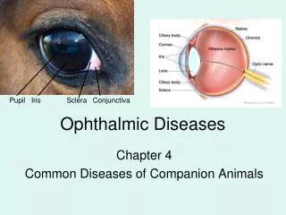
Visual pathway: • The optic nerve extends from the posterior pole of the eye (medially) to the optic chiasmawhere the nasal fibers cross to other side.After this characteristic crossing, the fibers of the optic nerve travel as the optic tractto the lateral geniculate body. then as optic radiation to the primary visual cortex. • The optic nerve has 2 sets of fibers: • 1-visual fibers going to lateral geniculate body • 2-afferent fibers of the pupillary reflex going to tectum of midbrain • the optic nerve is divided to 4 parts: • A-intraocular • B-orbital • C-intra canalicular • D-intracranial
Intraocular Portion of the Optic Nerve • The intraocular portion of the optic nerve is visible on ophthalmoscope as the • optic disc. All the retinal nerve fibers merge into the optic nerve here, and the central retinal vessels enter and leave the eye here. The complete absence of photoreceptors at this site creates a gap in the visual field known as the blind spot. • Shape and size: It is normally slightly vertically oval with an average area of 2.7mm2 and a horizontal diameter of 1.8mm. • Color: yellowish orange. • Margin: sharply defined and on the nasal side, the greater density of the nerve fibers makes the margin slightly less distinct than on the temporal side. • Optic cup: cavitation of the optic nerve, it’s the brightest part of the optic disc, no nerve fibers exit from it and there is a correlation between the size of it and the size of the optic disc. It is particularly important to document the size of the optic cup. This is specified as the horizontal and vertical ratios of cup to disc diameter (cup – disc ratio). normal ratio=0.4
Intraorbital and Intracranial Portion of the Optic Nerve The intraorbital portion begins after the nerve passes through a sieve-like plate of scleral connective tissue, the lamina cribrosa. After the optic nerve passes through the optic canal, the short intracranial portion begins and extends as far as the optic chiasm. Like the brain, the intraorbital and intracranial portions of the optic nerve are surrounded by sheaths of dura mater, pia mater, and arachnoid. The nerve receives its blood supply through the vascular pia mater sheath.
Papilledema: pathophysiology: As the optic nerve sheath is continuous with the subarachnoid space of the brain (and is regarded as an extension of the central nervous system), increased pressure is transmitted through to the optic nerve.Papilledema may be absent in cases of prior optic atrophy. blurred disc margins and dilated vessels . i.e optic disc swelling caused by increased intracranial pressure. Mostly bilateral.
Causes: • Increased intracranial pressure: • Brain tumor (60%) • Pseudotumor cerebri • Intracerebral hemorrahge • Venous sinus thrombosis • Maningitis/encephalitis • hydrocephalus • Others: • Malignant hypertension • Drugs:steriodstetracyclinelithiumnalidixic acid
Clinically: • Visual symptoms rare: • Some patients experience transient visual obscurations (inability to see in a particular part of the visual field for a period of time),graying-out of their vision, usually both eyes, especially when rising from a lying or sitting position, or transient flickering as if rapidly toggling a light switch). • Blurring of vision, constriction of the visual field, and decreased color perception may occur. • Diplopia,Visual acuity may be well-preserved, except in very advanced disease • History: • Headache(wosre on awakening and excacerbated by valsalva manuevers) • Nausea and vomiting • Palsatile tinnitus
Physical examination: *early: disc hyperemia, Subtle edema of the nerve fiber layer(nasaly) , Small hemorrhages of the nerve fiber, loss of venous pulsation *Late: elevated disc with blurred margins, Venous congestion develops, and peripapillary hemorrhages (over or adjacent to the disc) , exudates and cotton-wool spots. Paton’s lines(radial retinal lines cascading from the disc) ,choroidal folds *Chronic:disc hyperemia slowly subsides, giving way to a gray or pale disc that loses its central cup. may develop small glistening crystalline deposits (disc pseudodrusen).*On visual field examination, the physician may elicit an enlarged blind spot; the visual acuity may remain relatively intact until papilledema is severe or prolonged.Elevated grey optic disc with blurred margin,redial retinal hemorrhage,white-grey exudate.
Workup: Neuroimaging: (after fundoscopy) CT scan or MRI (with contrast)brain mass Magnetic resonance venographyvenous sinus thrombosis B-scan ultrasonography may be useful to rule out buried disc drusen. Fluorescein angiography Acute papilledema exhibits increased dilation of the peripapillary capillaries with late leakage of the dye. Lumber puncture: should be performed following a normal MRI to assess the opening pressure of the CSF and to obtain CSF for analysis to rule out neoplastic and infectious etiologies. It may provide some therapeutic benefit, as the CSF pressure is reduced temporarily
Treatment: Intracranial pressure should be reduced by treating the underlying disorder. Once intracranial pressure has been normalized, the pupillary edema will resolve within few weeks(6-8). Optic atrophy usually remains. prognosis depends on the degree of severity. • reduce the ICP: 1-medications(acetazolamide) 2-ventriculoperitoneal shunting 3-optic nerve decompression • Neurosurgery is required for space occupying lesions (i.e. tumors)
Optic neuritis: • i.e Demyelinating inflammation of the optic nerve . • Many cases are associated with multiple sclerosis. • It can be also isolated ON or associated with neuromyelitis optica(NMO) • The term papillitis is used if the optic nerve head is affected and retrobulbar neuritis if the nerve is affected more posteriorly. • Epidemiology: age:20-45, gender:females are twise as males, race: caucasians
History: • Sudden loss of vision (partial or complete).usually it is central vision(macular loss of vision) • Sudden blurred vision. • Vision loss may be exacerbated by heat or exercise(Uhthoff phenomenon). • Dyschromatopsia (change in color perception) in the affected eye occasionally may be more prominent than the decreased vision. • Retroorbital or ocular pain, usually exacerbated by eye movement and precedes the visual loss. • Frontal headache and tenderness of the globe. • -preceding history of viral illnes(40-70%have other symptoms) • -Objects moving in a straight line may appear to have a curved trajectory (Pulfrich phenomenon)
Physical examination: 1- relative afferent pupillary defect (RAPD) 2- varying degrees of reduction in vision 3-One third of patients with ON have a swollen disc (papillitis) 4- Reduced color vision 5- Central scotoma on field testing Investigation: 1-blood test 2-CSF:presence of myelin basic protein, oligoclonal bands, and elevated IgG index and synthesis rate in the CSF supports the diagnosis of MS 3-MRI: *a highly sensitive and specific tool in assessing inflammatory changes in the optic nerves, and helps to rule out structural lesions. *Utilization of fat saturation techniques helps to visualize gadolinium enhancement of the optic nerve and is the best imaging technique to visualize inflammation of the optic nerve *an MRI of the spinal cord is indicated in patients with suspected neuromyelitis optica (NMO).
VIDEO: RAPD RAPD:seen in: 1-optic nerve disease (ON, AION) 2-extensive retinal disease( maculopathy,RVO,RAO) #is usually because of asymmetric damage to the optic nerve or chiasm (less signal is transmitted along the afferent papilomotor pathway) One pupil dilates and the other constricts when light is swung from eye to eye #Causes: amblyopia , anisochorea(in epidural hematoma), dense blood (complete hyphema ,dense vitreal hemorrhage) but cataract doesn't
Treatment: When the presenting visual loss is mild, treatment is unnecessary. When visual acuity within the first week of onset is worse than 6/12, treatment may speed up recovery by several weeks. Regimen: Intravenous methylprednisolone sodium succinate (1 g daily) for 3 days followed by 11 days of oral prednisolone. Prognosis: -Visual acuity recovers well in most individuals. -The cumulative probability of developing MS by 15 years after onset of ON is 50% and strongly related to presence of lesions on a baseline MRI scan of the brain. Twenty-five percent of patients with no lesions on baseline brain MRI developed MS during follow-up compared with 72% of patients with one or more lesions
Anterior ischemic optic neuropathy Blood supply to the eye : internal carotid a. gives: **Ophthalmic artery gives off the following branches 1- Orbital group : Lacrimal artery Supraorbital artery Posterior ethmoidal artery Anterior ethmoidal artery Internal palpebral artery Frontal artery Nasal artery 2-Ocular group : Long ciliary artery Short ciliary artery Anterior ciliary artery Posterior ciliary artery Central retinal artery Muscular artery
Classification: Ischemic optic neuropathy is classified into : 1 – anterior ischemic optic neuropathy ( AION ) 2 – posterior ischemic optic neuropathy ( PION ) **AION is divided into : a-Arteric b-non-arteric **PION is divided into : a-Arteric b-non-arteric c-surgical PIONs
Non – arteric AION • most common type • Due to Ischemia in (anterior cilliary artery) circulation Etiologies : 1-Due to transient nonperfusion or hypoperfusion of the optic nerve head circulation . In most cases it is due to transient fall in BP, most commonly during sleep. In most cases there is no evidence of thromboembolic cause. In NA-AION, 73% of patients gave a definite history of discovering the visual loss on waking up . Patients who take anti hypertensive drugs are susceptible. 2 – emboli in minority of cases but it causes more damage
Risk factors: 1. Systemic risk factors: -arterial hypertension, nocturnal arterial hypotension, -diabetes mellitus, -ischemic heart disease, -hyperlipidemia, -atherosclerosis and arteriosclerosis -Other associated systemic diseases have also been reported, including: *sleep apnea, *arterial hypotension secondary to drugs, *malignant arterial hypertension *migraine.
2.Ocular and optic nerve head risk factors: -absent or small cup in the optic disc( cause crowded fibers as they exit from bruch membrane and the sclera , so ischemia will cause edema and this will cause swelling which will compress the capillaries and cause further ischemia) -angle closure glaucoma or any markedly raised IOP -marked optic disc edema due to any cause, -location of the watershed zone of the posterior ciliary arteries in relation to the optic disc, -optic disc drusen -cataract extraction. -Defective autoregulation of the optic nerve head may also play a role
Clinical features 1-Sudden painless loss of vision usually upon wakening 2-progressive visual loss can occur , the patients again usually notice it on waking in the morning. 3-NA-AION patients often complain of loss of vision towards the nose and less commonly altitudinal loss. 4-Later on, photophobia is a common complaint, particularly in bilateral cases. 5-Poor visual acuity but normal visual acuity doesn’t exclude NA – AION 6-Visual field loss is universal, so perimetry is very important to evaluate for visual loss loss of a hemifield (either the upper or lower half of the visual field, but not both) altitudinal visual defect. **Different visual field defect but combination of a relative inferior altitudinal defect with absolute inferior nasal defect is the most common pattern in NA-AION .
Right eye field of vision shows loss of inner and lower field of vision toward the nose. altitudinal visual feild defect
Right fundus photograph showing optic disc edema and hyperemia, with a splinter hemorrhage (arrow) during the acute phase of NA-AION, NO cupping
~ Managements of NA - AION~ Steroids : • possible mechanism : The faster resolution of optic disc edema with corticosteroid therapy compared to the untreated patients leads to progressive decrease of compression of the capillaries in the optic nerve head which leads to better blood flow in the capillaries and improved circulation in the optic nerve head improved function of the surviving but not functioning hypoxic axons Note: Aspirin is no beneficial because this is hypotensive event not thrombotic event Note: no evidence so far that intravitreal triamcinolone or anti-VEGF therapies have any beneficial effect .They may be harmful because of increase in IOP during injection.
Misconception about NA-AION • That NA-AION and cerebral stroke are similar in nature • That absence of optic disc cup is the main cause of development of NA-AION ( secondary contributing factor ) • That there is no spontaneous visual improvement in NA-AION( some studies shows improvement ) • That NA-AION is not seen in young persons • That all eyes with NA-AION initially have pale optic disc edema. Disc pallor actually starts to develop only 2 to 3 weeks after the onset of visual loss; before that there is no pale optic disc edema. • That inferior altitudinal defect is the classical diagnostic visual field defect in NA-AION. the most common defect in NA-AION eyes is the inferior nasal field defect. • That all eyes with NA-AION have poor visual acuity at onset. in eyes seen within 2 weeks of onset, 33% had 20/20 or better visual acuity. • That steroid therapy has no role in the management of NA-AION ( steroids improve visual acuity ) • That smoking is a risk factor for development of NA-AION. Two large prospective studies have shown that this is not true • That aspirin reduces the risk of second eye involvement by NA-AION • That all patients with NA-AION should be investigated for thrombophilia. As discussed above, NA-AION is not a thromboembolic disorder in the vast majority of cases
~ Arteritic -AION~ >>It is almost always due to Giant cell arteritis Other possible causes ( herpes , PAN, SLE ) • Giant cell arteritis (GCA) -Vasculitis affecting mainly medium and large arteries -Also called Temporal arteritis -Twice more common in females -Most patients are older than 50 , peak of incidence 60 – 80 -Incidence in USA is 0.5-27 cases per 100,000 people aged 50 years or older -Closely associated with PMR ( polymyalgia rheumatica )
Diagnostic criteria: Presence of three or more of the following is required for diagnose GCA : 1-Age 50 years or older 2-Newly onset localized headache 3-Temporal artery tenderness or decreased temporal artery pulse 4-ESR of at least 50 mm/h ( normally <= 17 mm/h) ( note : normal ESR doesn’t exclude , using both ESR and CPR is more specific ) 5-Abnormal artery biopsy specimen characterized by mononuclear infiltration or granulomatous inflammation Since it is vasculitis there are wide spectrum of manifestation include : -Constitutional ( fever , malaise , weight loss ) -Myalgia and arthralgia -Headache (extracranial, dull, boring, and burning , usually temporal ) and scalp tenderness -Jaw claudication and pain
Ophthalmic manifestation 1-Visual acuity: Although usually there is a marked deterioration of visual acuity in giant cell arteritis, almost normal visual acuity does not rule it out, and it is usually due to A-AION (affected central retinal artery) 2-Visual Fields: Compared to NA-AION, the visual defects are much more extensive and severe in A-AION. 3-Anterior segment of the eye: Usually it is normal except for relative afferent pupillary defect in unilateral A-AION cases. In an occasional case, there may be signs of anterior segment ischemia, with ocular hypotony, and/or marked exudation in the anterior chamber (erroneously diagnosed as anterior uveitis 4-Extraocular motility disorders: When present, this results in diplopia 5-Optic disc changes:Optic disc swelling, compared to NA-AION, usually has a diagnostic appearance in A-AION, i.e. chalky white color (seen in 69%). When optic disc edema resolves, the optic disc in the vast majority shows cupping indistinguishable from that seen in glaucomatous optic neuropathy , except that the disc rim is pale whereas it is of normal color in glaucomatous optic neuropathy. By contrast, in NA-AION no such cupping of the optic disc is seen
Fundus photographs of right eye with A-AION: (A) Before developing A-AION, (B) one week after developing A-AION with chalky white optic disc edema and (C) 4 months later showing optic disc cupping with a cup/disc ratio of 0.8
Management -High dose of steroids -Other immunosuppressant may be used ( cyclosporine , MTX , azathioprine ) -Sequential ESR determination may assist in determining the success of the high-dose corticosteroid therapy Differential Diagnoses: -Polymyalgia rheumatica-Transient ischemic attack-Neoplasms-Systemic infections-Amyloidosis with prominent vascular involvement-Arteriosclerotic vascular disease-Arteriovenous fistulas-Other forms of vasculitis
Idiopathic Intracranial Hypertension (IIH) -Also called Pseudotumor cerebri , Benign intracranial Hypertension -Increase in ICP with no lesion in the brain May be due to decrease absorption in arachinoid villi -Incidence in USA is .9 – 1 / 100,000 -Incidence in overweight is 7.9 – 20 / 100,000 ->90% of patients are women at childbearing age -Men are twice more likely to lose vision due to papilledema - OCP is a risk factor.
Clinical manifestation Patients usually complain of headache (worse in the morning and increased by valsalva maneuver ), diplopia Nonspecific symptoms may include dizziness, nausea, vomiting, photopsias, retrobulbar pain, and pulse-synchronous tinnitus. Transient visual obscurations: This visual symptom occurs in most patients. The disturbance can last up to 30 seconds and is described as a graying out of vision on a monocular or binocular basis. Orthostatic changes, such as standing up or bending over, can induce this symptom. Normal brain imaging , bilateral papilledema and increase in ICP No external ophthalmoplegia except for 6th nerve palsy
The most significant finding in patients with this disease is bilateral disc edema secondary to the increased intracranial pressure. • This papilledema varies from patient to patient and is indistinguishable from optic nerve swelling caused by intracranial space-occupying lesions. In more pronounced cases of disc swelling, macular involvement with subsequent edema and diminished central vision may be present. High-grade and atrophic papilledema in addition to subretinal hemorrhages are poor visual prognostic signs. • In some instances, the disc swelling is asymmetric, or, rarely, the appearance of the optic nerve may be relatively normal. • If left untreated, chronic disc swelling eventually leads to clinically significant visual loss. Although all patients present with enlarged blind spots during their initial perimetry, uncontrolled papilledema results in progressive peripheral visual field constriction or nerve fiber bundle defects (eg, nasal depression, nasal steps, arcuate scotomas). • The central visual field is affected in end-stage chronic papilledema. • The diplopia noted in patients with idiopathic intracranial hypertension is invariably due to unilateral or bilateral sixth nerve palsy. These cranial nerve palsies diminish with the lowering of the intracranial pressure.
Treatment: Weight reduction remove possible precipitating agent acetazolamide may decrease the ICP If not useful you may need to do VP shunt