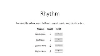
Rhythm Strips
190 likes | 364 Vues
Rhythm Strips. Jessica Wagner UMSON. EKG Grid. Ventricular Rate. 1 st method: Count the number of QRS complexes over the 6 second and then multiply by 10.

Rhythm Strips
E N D
Presentation Transcript
Rhythm Strips Jessica Wagner UMSON
Ventricular Rate 1st method: Count the number of QRS complexes over the 6 second and then multiply by 10 The 2ndmethod uses the small boxes. Count the number of small boxes for a typical R-R interval. Divide this number into 1500 to determine heart rate. In the second image, the number of small boxes for the R-R interval is 22.5. The heart rate is 1500/21.5, which is 69.8.
Ventricular Fibrillation • Fibrillation is an uncontrolled twitching or quivering of muscle fibers (fibrils). When it occurs in the lower chambers of the heart, it is called ventricular fibrillation • During VF, blood is not pumped from the heart Sudden cardiac death results • NO pulse http://health.nytimes.com/health/guides/disease/ventricular-fibrillation/overview.html
Causes of VF • Conditions that can lead to VF include: • Ischemia of the heart muscle • Electrocution accidents or injury to the heart • Heart attack (#1 cause!) • Heart disease that is present at birth (congenital) • Heart muscle disease, including cardiomyopathies • Heart surgery • Narrowed coronary arteries • Sudden cardiac death (commotiocordis), typically occurring in athletes after an injury over the surface of the heart
Risk Factors • Smoking • Diabetes • Hypertension
Symptoms • A person who has a VF episode can suddenly collapse or become unconscious, because the brain and muscles have stopped receiving blood from the heart. • The following symptoms may occur within minutes to 1 hour before the collapse: • Chest pain • Dizziness • Nausea • Rapid heartbeat • Shortness of breath
Diagnosing V Fib • NEVER from a 12-lead EKG b/c it will look the same as asytole • Cardiac monitor will show a very disorganized heart rhythm • Go in the room and check on your patient (pulses?) • “There is nothing to measure because your patient is dead… you better be doing CPR”
Treatment • VF is a medical EMERGENCY! TX STAT or death • If person has VF episode at home: 911 • Place person’s head and neck in line with the rest of their body to make breathing easier • Start CPR by doing chest compressions • Continue until person is alert or help arrives
Treatment • (#1) VF is treated by delivering a quick electric shock through the chest using a device called an external defibrillator • The electric shock can immediately restore the heartbeat to a normal rhythm, and should be done as quickly as possible • Many public places now have these machines (AED)
Treatment • An implantable cardioverter defibrillator (ICD) is a device that can be implanted in the chest wall of people who are at risk for this serious rhythm disorder • The ICD can help prevent sudden cardiac death by quickly sending an electrical shock when ventricular fibrillation occurs • Medicinesmay be given to control the heartbeat and heart function after 3 defibrillation attempts (i.e. amiodarone, vasopressin, epinepherine) • Antiarrhythmics agents: raise fibrillation and defibrillation threshold • Anticholinergic agents: reduce vagal activity improves conduction • Vasopressor agents: augment blood flow