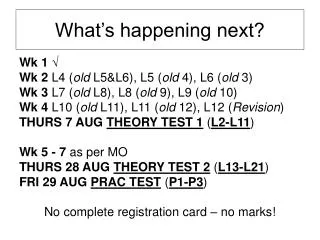
What’s happening next?
360 likes | 425 Vues
Refresh your knowledge about impulse transmission in sensory neurons, including receptor potentials, action potentials, and saltatory conduction. Learn about sensory signals, neuron structures, and connections with motor neurons. Prepare for upcoming theory and practical tests.

What’s happening next?
E N D
Presentation Transcript
What’s happening next? Wk 1√ Wk 2 L4 (old L5&L6), L5 (old 4), L6 (old 3) Wk 3 L7 (old L8), L8 (old 9), L9 (old 10) Wk 4 L10 (old L11), L11 (old 12), L12 (Revision) THURS 7 AUGTHEORY TEST 1 (L2-L11) Wk 5- 7 as per MO THURS 28 AUGTHEORY TEST 2 (L13-L21) FRI 29 AUGPRAC TEST (P1-P3) No complete registration card – no marks!
MBS 221Lecture 4: Refreshing prior knowledge about impulse transmission: mechanisms and influencesDr R. McBrideDepartment of Medical BiosciencesUniversity of the Western CapeOffice number B5.1(021) 959-2333rmcbride@uwc.ac.zaConsultation hrs: Wed 11h00 – 13h00
The stretch/knee-jerk reflex – the simple version… www.faculty.washington.edu
Sensory signals I • Recall that sensory receptors can either be: • discrete, individual cells (e.g. hair cells of the auditory and vestibular system, gustatory cells, rods and cones) or • sensory neurons with modified dendritic endings (e.g. nociceptors, olfactory neurons, Pacinian corpuscles, muscle spindles)
sensory receptors have three functional regions: • receptor region, containing stimulus-gated (mechanically-gated in the case of the muscle spindle) ion channels • spike-generating region, containing voltage-gated ion channels - this region may be anatomically separate from or continuous with the receptor region or with the axon itself 3. conducting region (axon), containing voltage-gated ion channels 1 2 3 3
recall that the modified dendritic endings of the muscle spindle wrap around intrafusal muscle fibers encapsulated within the belly of skeletal muscle and are sensitive to the: - length of the muscle • rate of change of length • stimulus-gated channels are located in the plasmalemma (in the receptor region of the sensory neuron) - these are cation-selective ion channels interconnected with each other and other cytoskeletal elements by cytoskeletal strands of spectrin
How does the stretching of the quadriceps (extensors) cause a receptor potential in the receptor region of the sensory neuron innervating that same (homonymous) muscle? -stimulus-gated channels in the plasmalemma of the receptor region open when the extensors stretch/lengthen • the influx of Na+ and/or Ca2+ depolarizes the membrane in this region • signal transduction occurs: mechanical energy electrical energy • the depolarization is called a receptor potential • this local or non-propagated potential is graded: • as the stimulus strength (amount of stretch) increases, so the amplitude (size) of the receptor potential increases – why? • because as the amount of stretch increases, so the time duration that ion channels are open increases
Recall that: - the cell bodies of sensory neurons cluster together outside the spinal cord in dorsal root ganglia - sensory neurons are pseudo-unipolar neurons • one axonal branch innervates the flexors = peripheral branch • one axonal branch extends into the dorsal horn of the spinal cord = central branch - the peripheral branch ends as the muscle spindle
How does the receptor potential cause an action potential (AP) in the spike-generating region of the sensory neuron? • as the membrane of the receptor region doesn’t contain voltage-gated ion channels, this region is electrically unexcitable and doesn’t generate APs. Why would this be beneficial? • if the receptor region generated APs, the graded nature of the receptor potential would be destroyed (recall the all-or-none phenomenon of the AP?) and the membrane potential would no longer encode stimulus intensity i.e. the nervous system wouldn’t be able to discern to what degree the extensors were being stretched • the receptor potential is passively propagated via continuous conduction to the spike-generating region/trigger zone/first node of Ranvier where voltage-gated Na+ channels are concentrated • take note that in sensory neurons, voltage-gated Na+ channels are concentrated at the first node of Ranvier, whereas in interneurons and motor neurons they are concentrated at the axon hillock • if the receptor potential is sufficiently large enough to bring the membrane potential of the spike-generating region to threshold, it will generate an AP • the AP is then actively propagated along the conducting region of the sensory neuron (myelinated axon) via saltatory conduction
Saltatory conduction Continuous conduction
Recall that the central branch of the sensory neuron forms excitatory connections with motor neurons that innervate the quadriceps and control their contraction
www.mona.uwi.edu • A single molecule forms the glutamate receptor and ion channel: • - are ionotropic i.e. have an immediate affect • are permeable to Na+ and K+ • are either NMDA or non-NMDA (kainate and AMPA) receptors
How does an AP arriving the sensory neuron terminal cause an AP in the motor neuron? • once an AP has been propagated along the conducting region (myelinated axon) of the sensory neuron via saltatory conduction and reaches the presynaptic terminal, it causes the opening of Ca2+ channels and the subsequent release of the neurotransmitter glutamate via exocytosis • the frequency and duration of arriving APs determines the amount of glutamate released • the binding of glutamate to ionotropic receptors on the postsynaptic membrane opens up voltage-gated channels via G-proteins and second messenger cascades • the net influx of Na+ into the postsynaptic membrane causes depolarization i.e. excitation • this local or unpropagated depolarization is called an excitatory postsynaptic potential (EPSP)
How does an AP arriving the sensory neuron terminal cause an AP in the motor neuron cont.? • like the receptor potential, the EPSP is a graded potential but it spreads passively to the axon hillock of the motor neuron • only if the EPSP is sufficiently large enough to bring the membrane potential of the axon hillock to threshold, it will generate an AP • if the strength of the stretch increases, more sensory neurons will be excited and the depolarization produced by the EPSP will be large enough to reach threshold • the AP is then actively propagated along the myelinated sensory axon via saltatory conduction
Recall the features of the nerve-muscle synapse or neuromuscular junction (NMJ): - directly gated transmission - motor unit - motor end plate - presynaptic boutons - synaptic cleft - active zones - synaptic vesicles containing acetylcholine (ACh) - mitochondria - voltage-gated Ca2+ channels - junctional folds - ACh-gated channels - voltage-gated Na+ channels - basement membrane with acetylcholinesterase
Above, ACh receptors at the top one-third of the postsynaptic junctional folds can be seen in black. Can you see the features of striated muscle at the bottom of the picture?
A single molecule forms the ACh receptor and ion channel: - called a nicotinic acetylcholine receptor (nAChR) because it binds nicotine - nAChRs are ionotropic i.e. have an immediate affect - are permeable to Na+, K+ and Ca2+ - muscle and neuronal type
How does the action potential (AP) arriving at the motor neuron terminal stimulate/activate skeletal muscle? i.e. how do the events at the NMJ cause skeletal muscle contraction? • the motor neuron excites the muscle by opening nAChRs at the motor end-plate • the net influx of Na+ into the motor end-plate causes depolarization i.e. excitation • the excitatory postsynaptic potential (EPSP) is called the end-plate potential (a local/graded potential) • the amplitude (size) of the end-plate potential is large enough (70 mV) to rapidly activate the voltage-gated Na+ channels in the junctional folds • the further influx of Na+ into the motor end-plate converts the end-plate potential into an AP • the AP is then propagated (spread) along the length of the muscle fiber • take note that in the CNS, EPSPs normally have a very small amplitude (< 1 mV), and so input from many presynaptic neurons is needed to generate APs (spatial summation)
How does the action potential (AP) arriving at the motor neuron terminal stimulate/activate skeletal muscle cont.? i.e. how do the events at the NMJ cause skeletal muscle contraction? - the AP in the muscle fibers opens up Ca2+channels in the sarcoplasmic reticulum, Ca2+binds to troponin, troponin-tropomyosin conformation changes, myosin binds to actin, cross-bridges shorten and muscle contracts!
Will the arrival of an AP at the motor neuron terminal ever directly inhibit skeletal muscle?i.e. will it ever directly cause skeletal muscle relaxation? • No! the binding of ACh to nAChRs will always have a stimulatory/excitatory effect on skeletal muscle • but there are certain diseases or toxin exposures that result in skeletal muscle relaxation. Can you think of any?
Synaptic transmission between central neurons is more complex than synaptic transmission at the NMJ: • many hundreds of sensory neurons converge on one motor neuron (recall convergent, divergent and osscilating pathways) • motor neurons receive both excitatory and inhibitory inputs and these signals need to be integrated through temporal and spatial summation • more than one neurotransmitter type is released at and more than one receptor type are activated at central synapses • motor neurons have both ionotropic and metabotropic receptors (and the second messengers they activate) • connections between pre- and postsynaptic neurons are only modestly effective • 50-100 excitatory neurons need to fire simultaneously in order for the EPSP to reach threshold and an AP to be triggered in the motor neuron
Possible test/exam questions from this lecture… unless otherwise indicated: • all scanned images are from Kandel ER, Schwartz JH and Jessell TM. Principles of Neural Science. 4th International Edition. McGraw-Hill Companies, Inc. • Define/describe the following terms/concepts (2-5 marks): • receptor, spike-generating and conducting region of receptors, stimulus and voltage-gated ion channels, spectrin, receptor potential, dorsal root ganglion, peripheral and central branch of pseudo-unipolar neuron, continuous and saltatory conduction, glutamate, ionotropic glutamate receptors, excitatory postsynaptic potential (EPSP), neuromuscular junction (NMJ), nicotinic acetylcholine receptors (nAChRs), end-plate potential
Possible test/exam questions from this lecture… • Answer the following questions (5-10 marks): • name and describe the three functional regions of receptors • explain how the stretching of the quadriceps (extensors) causes a receptor potential in the receptor region of the sensory neuron innervating that same (homonymous) muscle • explain how the receptor potential causes an action potential in the spike-generating region of the sensory neuron • compare the propagation of receptor and action potentials in sensory neurons • explain how an action potential arriving at the sensory neuron terminal causes an action potential in the motor neuron • explain how an action potential arriving at the motor neuron terminal stimulates/activates skeletal muscle • explain why the arrival of an action potential at the motor neuron terminal will never ever directly inhibit skeletal muscle • discuss the complexities of synaptic transmission between central neurons compared to synaptic transmission at the NMJ