Download
1 / 5
50 likes | 60 Vues
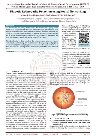
Diabetic Retinopathy is disease of eye that causes injury to the retina and it may ultimately lead to absolute loss of sight. Tests which detect the diabetic retinopathy like visual acuity examination, pupil dilation, and Optical Coherence Tomography OCT are time intense and affects patients too. Fuzzy inference based model allows intelligent system to examine the patient and then infers a conclusion. The proposed fuzzy expert model deploys 9 Input Parameters Intra ocular pressure, visual field, glucose level in blood, high density lipoprotein, low density lipoprotein, hemoglobin, Glycated sugar, blood pressure and Triglyceride . In this model, expert system recognizes the normal eye, diabetic retinopathy Severe , diabetic retinopathy moderate and diabetic retinopathy Mild on the behalf of these 9 Parameters. This technique has efficient low computational cost and has comparative outcomes to those of the ophthalmologist having 88 accuracy. Ratish | Neeru Malhotra | Vishav Kapoor "Fuzzy Based Decision Making System for the Detection of Diabetic Retinopathy" Published in International Journal of Trend in Scientific Research and Development (ijtsrd), ISSN: 2456-6470, Volume-4 | Issue-5 , August 2020, URL: https://www.ijtsrd.com/papers/ijtsrd30325.pdf Paper Url :https://www.ijtsrd.com/engineering/electronics-and-communication-engineering/30325/fuzzy-based-decision-making-system-for-the-detection-of-diabetic-retinopathy/ratish<br>

E N D
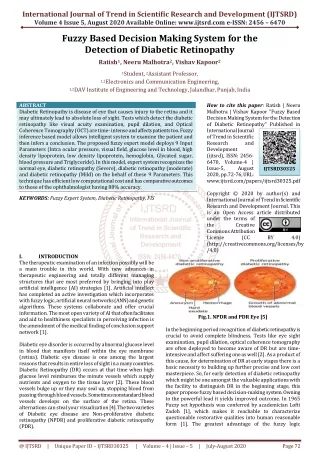
International Journal of Trend in Scientific Research and Development (IJTSRD) Volume 4 Issue 5, August 2020 Available Online: www.ijtsrd.com e-ISSN: 2456 – 6470 Fuzzy Based Decision Making System for the Detection of Diabetic Retinopathy Ratish1, Neeru Malhotra2, Vishav Kapoor2 1Student, 2Assistant Professor, 1,2Electronics and Communication Engineering, 1,2DAV Institute of Engineering and Technology, Jalandhar, Punjab, India ABSTRACT Diabetic Retinopathy is disease of eye that causes injury to the retina and it may ultimately lead to absolute loss of sight. Tests which detect the diabetic retinopathy like visual acuity examination, pupil dilation, and Optical Coherence Tomography (OCT) are time- intense and affects patients too. Fuzzy inference based model allows intelligent system to examine the patient and then infers a conclusion. The proposed fuzzy expert model deploys 9 Input Parameters (Intra ocular pressure, visual field, glucose level in blood, high density lipoprotein, low density lipoprotein, hemoglobin, Glycated sugar, blood pressure and Triglyceride). In this model, expert system recognizes the normal eye, diabetic retinopathy (Severe), diabetic retinopathy (moderate) and diabetic retinopathy (Mild) on the behalf of these 9 Parameters. This technique has efficient low computational cost and has comparative outcomes to those of the ophthalmologist having 88% accuracy. KEYWORDS: Fuzzy Expert System, Diabetic Retinopathy, FIS How to cite this paper: Ratish | Neeru Malhotra | Vishav Kapoor "Fuzzy Based Decision Making System for the Detection of Diabetic Retinopathy" Published in International Journal of Trend in Scientific Research and Development (ijtsrd), ISSN: 2456- 6470, Volume-4 | Issue-5, August 2020, pp.72-76, URL: www.ijtsrd.com/papers/ijtsrd30325.pdf Copyright © 2020 by author(s) and International Journal of Trend in Scientific Research and Development Journal. This is an Open Access article distributed under the terms of the Creative Commons Attribution License (CC (http://creativecommons.org/licenses/by /4.0) IJTSRD30325 BY 4.0) I. The therapeutic examination of an infection possibly will be a main trouble in this world. With new advances in therapeutic engineering and totally different managing structures that are most preferred by bringing into play artificial intelligence (AI) strategies [1]. Artificial intellect has completed an active investigation which incorporates with fuzzy logic, artificial neural networks (ANN) and genetic algorithms. These systems collaborate and offer crucial information. The most open variety of AI that often facilitates and aid to healthiness specialists in perceiving infection is the amendment of the medical finding of conclusion support network [1]. Diabetic eye disorder is occurred by abnormal glucose level in blood that manifests itself within the eye membrane (retina). Diabetic eye disease is one among the largest reasons that results in entire loss of sight in a many countries. Diabetic Retinopathy (DR) occurs at that time when high glucose level reimburses the minute vessels which supply nutrients and oxygen to the tissue layer [2]. These blood vessels bulge up or they may seal up, stopping blood from passing through blood vessels. Sometimes nonstandard blood vessels develops on the surface of the retina. These alternations can steal your visualization [4]. The two varieties of Diabetic eye disease are Non-proliferative diabetic retinopathy (NPDR) and proliferative diabetic retinopathy (PDR). INTRODUCTION Fig.1. NPDR and PDR Eye [5] In the beginning period recognition of diabetic retinopathy is crucial to avoid complete blindness. Tests like eye sight examination, pupil dilation, optical coherence tomography are often deployed to become aware of DR but are time- intensive and affect suffering one as well [2]. As a product of this cause, for determination of DR at early stages there is a basic necessity to building up further precise and low cost masterpiece. So, for early detection of diabetic retinopathy which might be one amongst the valuable applications with the facility to distinguish DR in the beginning stage, this paper propose fuzzy based decision-making system. Owning to the powerful lead it yields improved outcome. In 1965 Fuzzy set hypothesis was conferred by academician Lofti Zadeh [1], which makes it reachable to characterize questionable restorative qualities into human reasonable form [1]. The greatest advantage of the fuzzy logic @ IJTSRD | Unique Paper ID – IJTSRD30325 | Volume – 4 | Issue – 5 | July-August 2020 Page 72
International Journal of Trend in Scientific Research and Development (IJTSRD) @ www.ijtsrd.com eISSN: 2456-6470 framework lies in the way that analysts can demonstrate dubious, complicated framework into uncomplicated human understandable form by utilizing human understanding and education as fuzzy guiding principle as locate of linguistic factors [1].This paper mentions a master structure by assembling fuzzy set judgments to infer diabetic retinopathy from its suggested indications. With medical specialist’s data, fuzzy standards are put forth for a bit more superior cognitive practice. For decision of diabetic retinopathy this paper provides information based on the expert structure. Moreover, laying out and implementing outcome for examination of diabetic retinopathy by using fuzzy intrusion system is organized. Fuzzy inference system is armed with at least 9 parameters of diabetic retinopathy. To utilize the therapeutic expert’s data for significant patient's symptoms and give an exact decision as per fuzzy guidelines are completed by the fuzzy rules based system. Fig.2. Fuzzy Expert System [1] II. RELATED WORK There is diversity of works and closely analyzed literature that demonstrate the accomplishment and representation of medical expert system. Furtado P. et al. (2017) introduced Segmentation by Density Clustering of Fundus Images (EFI) in Diabetic Retinopathy. By using a set of metrics: Runtime is the time segmentation takes to run, versus the number of super pixels, the purposed system compared the resulting segmentation. Author concluded that Simple Linear Iterative Clustering (SLIC) + Density-based spatial clustering of applications with noise (DBSCAN) isolate lesions better [3].Bhatia k. etal. (2016) proposed methods such as ANN, SVM etc. To distinguish the diabetic retinopathy among the diabetic patients a programmed coordination was developed which aimed to help ophthalmologists to become aware of indications of diabetic eye disease with effortlessness and also highlights various technologies which were used for recognition of diabetic eye disease [2].Kusakunniran w. et al. (2016) elaborated the procedures to the automatic retinal image quality assessment and hard exudates segmentation which was performed on tha basis of contrast histogram. Both optic disk (OD) and hard exudates segmentations are based lying on the image thresholding using iterative selection and the grabcut. Author concluded that the projected method has over 90% accuracy [13]. Labhade J. et al. (2016) introduced soft computing techniques (Support Vector Machine (SVM), Random Forests, Gradient boost, AdaBoost, Gaussian Naive Bayes) for DR detection and each provides different accuracy. Author concluded that the SVM classifier provides better testing accuracy up to 88% while random forests Technique and Gradient boost provides 83%. The Gaussian NB and AdaBoost classifier gave poor accuracy [14].Paing M. et al. (2016) projected a system for detecting and classifying DR using ANN. Many lesions namely blood vessels, exudates and micro-aneurysms from respective images were found by the authors who extracted necessary features and classified them with the help of ANN classifier. Author concluded that sensitivity, precision and accuracy of the system were 95%, 95%, 96% respectively [15]. Zohora S. et al. (2016) reviewed on various automated techniques by means of unlike algorithms which resulted in better performance for exudates detection. Author concluded that among the range of approaches used the act of fuzzy c-means clustering techniques detects exudates more accurately than other methods explained in exudates detection [16]. Ashe S. S. et al. (2016) projected algorithmic rule which is based on intensity based preprocessing which is fitting for the recognition of optic disc (OD) and Macula before the investigation of Diabetic Retinopathy. The two key factors which require to be localized for analyzing diabetic retinopathy are Optic Disc and Macula. The intensity primarily based on threshold selection a criterion was used for the detection. With their proposed algorithm OD and Macula section can detect effectively [9]. Ibraheem S. et al. (2015) proposed classification of the diabetic retinopathy into Exudates, micro-aneurysms and hemorrhages by using fundus images. A quantity of features such as mean, standard deviation, discrepancy, energy, Homogeneity and Entropy of the pre-processed images are extracted to describe the image contented to formulate a finding of the diabetic retinopathy. Author concluded that the categorization precision can provide a better result and Image processing techniques can condense the job of ophthalmologists [11]. Sangwan S. et al. (2015) proposed categorization of Normal, NPDR or PDR affected eye with high accuracy percentage of 92.6%. Author concluded that SVM will be utilized efficiently as classifiers for detecting observe interrelated diseases causes by diabetic [17]. Shojaeipour A. et al.(2014) projected a CAD system for the diagnosis of diabetic retinopathy by using digital retina images and classifying these images into two categories, diabetic retinopathy eyes and non-diabetic retinopathy eyes (healthy eyes). Author concluded that the qualifications of the system increased and the possibility of errors are reduced with each training epoch [7]. III. PROPOSED SYSTEM This part clarifies the approach embraced in building the general fuzzy framework for decision making framework. The fuzzy inference framework is a structure which is reliant on fuzzy set hypothesis, picks up a fuzzy representation of patient's manifestation and accordingly induces fuzzy relationship. With a specified ambition to realize fuzzy depiction to fullest i.e. interpretability and the capacity to handle generalization is very critical. The word oversimplification imply that capacity to express the state-activity as compact as feasible. Generalization rules allow extra strong rule base, fast induction and well defined fuzzy interpretability. A fuzzy dependent decision support system accomplishes master information and experience in comprehension of IF-ELSE guidelines to plan fuzzy inference. Hence, a fuzzy master framework permits a straightforward path for planning an Crisp Input Fuzzification Fuzzy Inference system Fuzzy rule Base Defuzzification Crisp output Values to accomplish elevated @ IJTSRD | Unique Paper ID – IJTSRD30325 | Volume – 4 | Issue – 5 | July-August 2020 Page 73
International Journal of Trend in Scientific Research and Development (IJTSRD) @ www.ijtsrd.com eISSN: 2456-6470 exact arrangement with help from an uncertain area. The given fuzzy set relating with a participation work characterizes the information credit to its exact membership and it must lie in range of (0, 1). A fuzzy set has no cotemporary worth and has a fuzzy intermediate. The quadrilateral membership plot is a task having four variables a, b, c, d where a and d denote feet of quad with relationship degree 0 and b and c be a symbol of shoulders of trapezoid with attachment degree 1. IV. METHODOLOGY In Fig. 4, for conniving the expert system nine contribution variables i.e. IOP, visual field, fasting blood sugar, HDL, LDL, HB, HBA1C, BP, and Triglyceride are used. These inputs are used infer the health standing of person. After selecting the participation variables consecutive step is to fuzzify the factors i.e. we have to establish the fuzzy sets for each and every contribution variable and the consequent vary of the belonging to each fuzzy set. Fuzzy rule-base allows experts knowledge to reflect on indications of enduring and then support the rules developed to give a precise decision. On- line primary medical care symptoms appraisal implies citing of these symptoms that are vital for the investigation and inference of disease. Figure 4.1 represent the methodology for the proposed system. Fuzzy Inference System (FIS) and Graphical User Interface (GUI) are very powerful tools provided by MATLAB to propose a Fuzzy verdict structure. The training part with reference to fuzzy inference system is done by the FIS editor, which is another strong tool provided by MATLAB. There is a straightforward and logical illustration in figure 4.2 that represent the denomination of all participation (9 input parameter values) on the left hand side and those of each output on the right hand side. Yet, the quantity of inputs could also be restricted by the presented memory of your machine. fuzzify the variables i.e. we have to decide the fuzzy sets for each one input variable and also the corresponding vary of the belonging to every fuzzy set. A.Cholestrol (HDL,LDL,Tryglyceride) B.General Health (Intraocular Pressure,Fasting blood Sugar,Visual Acuity, Blood Pressure) C.HB, HBA1C Fig.4.2. Hierarchical FIS with 9 inputs & 1 output B.Membership Functions Every single one association functions are connected with each variable. The relationship functions of parameters are shown below and do not depict the unambiguous outline of the membership functions. The membership function is used to edit rules and be evidence for all of the association functions for the integrated fuzzy inference system, together cooperating input and output parameters. Fig.4.3(a). Membership Plot for Intraocular Pressure Fig.4.3(b). Membership plot for fasting blood sugar It is the manner of the unification of the rules. The membership functions of the entire rules previously antecedently clipped at some stage in rule evaluation are in use and unified to one fuzzy set. In progression a quantity of clipped subsequent relationship functions are represented conjointly to one fuzzy set for each production variable. The Mamdani inference method is used for the regarding inference. C.Rule Editor Rule editor is used for editing the directory of rules that delineate the presentation of the structure. A comprised outsized editable text field is used to display, write the rules Fig.4.1. Methodology to Implement Proposed System A.Input Variables For manipulative the skilled system nine input multivariate i.e. IOP, visual field, fasting blood sugar, HDL, LDL, HB, HBA1C, BP, and Triglyceride are used. These inputs are used to diagnose the well being condition of the person. After choosing the participation variables successive stride is to @ IJTSRD | Unique Paper ID – IJTSRD30325 | Volume – 4 | Issue – 5 | July-August 2020 Page 74
International Journal of Trend in Scientific Research and Development (IJTSRD) @ www.ijtsrd.com eISSN: 2456-6470 and to edit the same. In addition, rule editor has various well-known with landmarks constant as those inside the FIS (fuzzy Inference system) editor and, along with the menu bar and also the status line membership function Editor. Table I represents result of nine Parameters are computed by proposed method for the test images and the Doctor’s Observation. It was found that all the Diabetic retinopathy images were classified as Diabetic retinopathy by Fuzzy classifier while out of 50 normal images, 6 were wrongly classified as Diabetic retinopathy. TABLE I. RESULTS Fig.4.4. Rule Editor D.Fuzzification and Defuzzification The next phase of fuzzy expert system is the Fuzzification. It is the technique of mapping a crisp assessment of an input to relationship degrees in several fuzzy linguistic multivariates. Defuzzification is the contrary course of action of Fuzzification. Thus crisp inference output is known by the Defuzzification method later than estimating its input significance. V. EXPERIMENTAL RESULTS A.Rule Viewer Rule observer to analysis the fuzzy inference system. Exercise this observer as an indicative to check, for instance, the entity membership function semblance implication the results. The rule viewer unveils the information of the complete fuzzy inference procedure. The status line and the menu bar intimate items are introduced additionally. To enter a specific input value, a text field is given at the lower right position. Figure 5.1 displays the rule viewer of the anticipated system. It indicates the outcome of entire proposed system. From the left plane at the peak we get defuzzified values, we get = 6.35 which means the person is normal. µA=Microaneurysms, H= hemorrhages, EX= exudates, NV= Neovascularization Accuracy is defined as the ratio of number of correct decisions to the total number of patients [29]. Accuracy = 46 x 100 = 92% 50 VI. CONCLUSION Diabetic retinopathy is most extensive disease today, so early recognition is exceptionally important to keep individuals experiencing Diabetic retinopathy. Early finding is the highest loyalty choice which gives medical doctors to separate Normal eye and Diabetic eye. In this research, we have exhibited a fuzzy structure on decision supportive network for the pronouncement of Diabetic retinopathy. The proposed fuzzy interference framework predicts the healthy vision and Diabetic eye. The anticipated approach can manage different sources of input which can be far superior to handle vulnerability during investigating period. The consequences of proposed system are compared with clinical dataset of 50 patients; the system gives promising result over 92% accuracy. This present framework can be extended by expanding number of inputs. The limitations of our system are Age factor and hereditary relations which cannot be considered in our system, Severity level cannot be inferred and Treatment cannot be suggested in our system. To make Android App for Diabetic retinopathy detection and to infer severity level of disease will be beneficial for Society. Therefore, This Technology will have a Great Impact in Future. REFERENCE [1]John A., Sharma A., Rehani V., “Glaucoma Detection System (GDS); a Novel GUI based Approach for Glaucoma Detection” International Journal of Fig.5.1. Rule Viewer B.Surface Viewer To evaluation the dependence of 1 of the outputs on 1 or 2 of the inputs Surface viewer is used, for the fuzzy inference system(FIS) it generate and design an output surface plot. From 2 input variables and one production variable of a FIS It engender a 3-d surface. Fig.5.2. Surface view of Intraocular Pressure and fasting blood sugar @ IJTSRD | Unique Paper ID – IJTSRD30325 | Volume – 4 | Issue – 5 | July-August 2020 Page 75
International Journal of Trend in Scientific Research and Development (IJTSRD) @ www.ijtsrd.com eISSN: 2456-6470 Computational Engineering Research (IJCER), vol.07 November, 2017. [2]Bhatia R., Arora S., Tomar R., “Diagnosis of Diabetic Retinopathy Using Machine Learning Classification Algorithm”, Proceedings of IEEE 2nd International Conference on Next Technologies (NGCT-2016). [3]Furtado P., Travassos C., Monteiro R., Oliveira S., Baptista C., Carrilho F., “Segmentation of Eye Fundus Images by Density Clustering in Diabetic Retinopathy”, Biomedical & Health Informatics (BHI), Proceedings of IEEE EMBS International Conference , 2017 . [4]Eye Smart - What Is Diabetic Retinopathy?, 2017 American Academy of Ophthalmology. [5]Maxivision eye hospital, “eye focus: understanding diabetic http://maxivisioneyehospital.wordpress.com/2013/0 7/03/eye-focus-undersatnd-diabetic-retinopathy. [6]Ahmad A., Mansoor A., Mumtaz R., Khan M., “Image Processing and Classification in Diabetic Retinopathy: A Review”, visual information processing (EUVIP), 5th European workshop IEEE, 2014. [7]Shojaeipour A., Nordin J., Hadavi N., “Using Image Processing Methods Retinopathy” , Proceedings of IEEE International Symposium on Robotics Automation, 2014. [8]Salam A., Akram M., Wazir K., Anwar S., “Autonomous Glaucoma Detection From Fundus Image Using Cup to Disc Ratio and Hybrid Features” International Symposium on Signal Processing and Information Technology, Proceedings of IEEE, 2015. [9]Ashe H., Ali I., “A New Method for Detection of Optical Disc and Macula for Diabetic Retinopathy”, Proceedings of IEEE 2nd International Communication, Control and Intelligent Systems (CCIS), 2016. [10]Dhanasekaran R., Mahendran G., Murugeswari S., Fargana S., “Investigation of Diabetic Retinopathy using GMM classifier”, Proceedings of IEEE International Conference on Advanced Communication Control and Computing Technologies (ICACCCT), 2016. [11]Ibraheem S., Khalaf A., Salah S., “Classification of Diabetic Retinopathy Types Using Fuzzy Logic”, International Journal of Advanced Research in Computer Science and Software Engineering, Volume 5, Issue 3, March, 2015. [12]Gupta V., Teja S., Gupta S. ,Sengar P, “Extraction of Blood Veins from the Fundus Image to Detect Diabetic Retinopathy”, Proceedings of IEEE 1st International Conference on Power Electronics Intelligent Control and Energy Systems (ICPEICES-2016). [13]Kusakunniran W., Rattanachoosin J., Sutassananon K., Anekkitphanich P., “Automatic Quality Assessment and Segmentation of Diabetic Retinopathy Images”, Proceedings of IEEE Region 10 Conference (TENCON), 2016. [14]Labhade J., Chouthmol L., Deshmukh S., “Diabetic Retinopathy Detection Techniques” , Proceedings of IEEE International Conference on Automatic Control and Dynamic Optimization Techniques (ICACDOT),2016. [15]Paing M., Choomchuay S., Yodprom R., “Detection of Lesions and Classification of Diabetic Retinopathy Using Fundus Images”, Proceedings of IEEE The Biomedical Engineering International Conference (BMEiCON-2016). [16]Zohora S.,Khan A., Chakraborty S., Dey N., “Detection of Exudates in Diabetic Retinopathy: A Review”, Proceedings of IEEE International Conference on Electrical, Electronics, and Optimization Techniques (ICEEOT), 2016. [17]Sangwan S., Sharma V., Kakkar M., “Identification of Different Stages of Diabetic Retinopathy”, Proceedings of IEEE International Conference on Computer and Computational Sciences (ICCCS). [18]Sreng S., Takada J., Maneerat N., Isarakorn D., “Feature Extraction from Retinal Fundus Image for Early Detection of Diabetic Retinopathy”, Proceedings of IEEE R10-HTC, 2013. [19]Prasad D., Vibha L., R V., “Early Detection of Diabetic Retinopathy from Digital Retinal Fundus Images”, Proceedings of IEEE Recent Advances in Intelligent Computational Systems (RAICS), 2015. [20]Gandhi M., “Investigation of Severity of Diabetic Retinopathy by detecting Exudates with respect to Macula”, Proceedings of IEEE ICCSP 2015 conference. [21]Khurana A. K., “Diseases of the retina,” in Comprehensive Ophthalmology, 5th ed. New Delhi, India: New Age International, 2012, ch. 11. [22]Mayoclinic.org, “Diseases and Conditions Diabetic retinopathy Basic Tests and Diagnosis”, 2015. [online]. Available:http://www.mayoclinic.org/diseases- conditions/diabetic-retinopathy/basics/tests- diagnosis/con-20023311. [Accessed: 7-July-2017]. [23]nei.nih.gov, “Healthy Eyes Eye Exam”, 2010.[online]. Available:https://nei.nih.gov/healthyeyes/eyeexam+W hat+is+a+comprehensive+dilated+eye+exam?. [Accessed: 6-July-2017]. [24]webmd.com, “cholesterol management tc lipid panel topic overview”, 2015. [online]. Available: http: //www. webmd. management/tc/lipid-panel-topic-overview. [Accessed: 7-July-2017]. [25]diabeticlivingonline.com, “monitoring abcs diabetes a1c blood pressure and cholestrol”, 2014. [online]. Available: diabeticlivingonline.com/monitoring/abcs-diabetes- abcs-diabetes-a1c-blood-pressure-and-cholesterol. [Accessed: 8-July-2017]. [26]www.aao.org, “eye health dignosis”,[online]. available//http:www.aao.org /eye- health/disease/glaucoma-diagnosis. [Accessed: 8-July- 2017]. [27]Lamani D., Kumar R., “Different Clinical Parameters to Diagnose Glaucoma Disease: A Review”International Journal of Computer Applications (0975 – 8887) Volume 116 – No. 23, April 2015. [28]www.google.co.in, “tonometry”, Available: //www.google.co.in/search?q=tonometry. [Accessed: 18-July-2017]. [29]A. Salam, M. Akram, K. Wazir, S.M. Anwar, M. Majid, “Autonomous Glaucoma Detection From Fundus Image Using Cup to Disc Ratio and Hybrid Features” proceeding of the IEEE International Symposium on Signal Processing and Information Technology, (2015) , pp: 370-374. Generation Computing retinopathy”, for Diagnosis Diabetic and Manufacturing Conference on com/cholesterol- http: //www. disease glaucoma 2015.[online]. https: Using Soft Computing @ IJTSRD | Unique Paper ID – IJTSRD30325 | Volume – 4 | Issue – 5 | July-August 2020 Page 76