Download

1 / 6
60 likes | 94 Vues
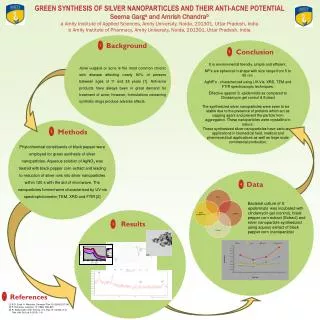
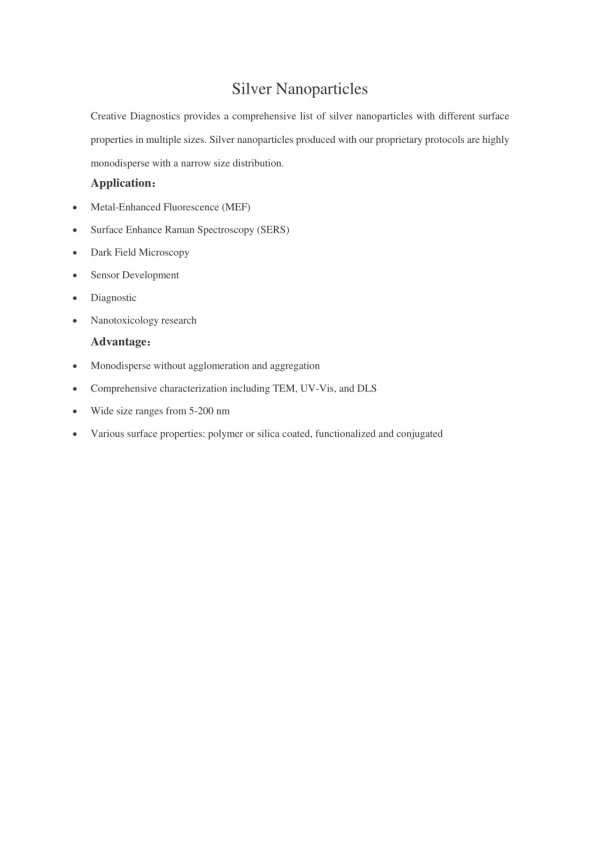
Nanosilver and other forms of silver are widely used nowadays for their antibacterial activity. The different forms of silver, including silver salts, silver oxides and silver materials appearing as silver wires, silver nanopartilces AgNP and others, which are used in consumer and medical products, may have different physic chemical properties such as distinct solubility and surface to volume ratio, which all may affect their fate and biological activity. AgNPs display different applications in the field of dentistry owing to their antimicrobial effect. Various evidences has been found for several advantages of using natural or synthetic organic nanostructures in a wide variety of dental fields, from implantology, endodontics and periodontics, to regenerative dentistry and wound healing. This review aims to discuss the current progress in this field, highlighting aspects regarding silver nanoparticles incorporation such as antimicrobial potential, mechanical properties and long term effectiveness. Chandani Desai | Siddharth Desai "Silver Nanoparticles in Dental Cure: Scenario and Confronts" Published in International Journal of Trend in Scientific Research and Development (ijtsrd), ISSN: 2456-6470, Volume-2 | Issue-2 , February 2018, URL: https://www.ijtsrd.com/papers/ijtsrd8319.pdf Paper URL: http://www.ijtsrd.com/biological-science/biotechnology/8319/silver-nanoparticles-in-dental-cure-scenario-and-confronts/chandani-desai<br>

E N D
International Journal of Trend in Scientific Research and Development (IJTSRD) International Open Access Journal ISSN No: 2456 - 6470 | www.ijtsrd.com | Volume - 2 | Issue – 2 Silver Nanoparticles in Dental Cure: Scenario and Confronts Chandani Desai Shree Ramkrishna Institute of Computer Education And Applied Sciences Surat-395007, Gujarat, India ABSTRACT Nanosilver and other forms of silver are widely used nowadays for their antibacterial activity. The different forms of silver, including silver salts, silver oxides and silver materials appearing as silver wires, silver nanopartilces (AgNP) and others, which are used in consumer and medical products, may have different physic-chemical properties such as distinct solubility and surface-to-volume ratio, which all may affect their fate and biological activity. AgNPs display different applications in the field of dentistry owing to their antimicrobial effect. Various evidences has been found for several advantages of using natural or synthetic organic nanostructures in a wide variety of dental fields, from implantology, endodontics and periodontics, to regenerative dentistry and wound healing. This review aims to discuss the current progress in this field, highlighting aspects regarding silver nanoparticles incorporation such as antimicrobial potential, mechanical properties and long-term effectiveness. Keywords: Silver Nanoparticles; Dentristry; Restorative Material; Periodontal Pockets; Implantology INTRODUCTION Siddharth Desai Prosthodontics Department, Vaidik Dental College, Hospital and Research Centre, Daman, India physics [1]. Over the years, silver compounds and NPs have exhibited antibacterial activity resulting in the widespread use of silver nanoparticles (Ag NPs) in bedding, washing machines, water purification, toothpaste, shampoo and rinse, nursing bottles, fabrics, deodorants, filters, kitchen utensils, toys and humidifiers [2]. Furthermore, silver compounds and NPs [3] have been studied for dental applications including dental restorative material [4], endodontic retrofill cement [5], dental implants [6] and caries inhibitory solution [7]. AgNPs have also been applied in several areas of dentistry, as endodontics [8, 9], dental prostheses [10], implantology [11, 12], and restorative dentistry [13–15]. AgNPs incorporation aims to avoid or at least to decrease the microbial colonization over dental materials, increasing oral health levels and improving Nanotechnology is emerging as an interdisciplinary field that is undergoing rapid development and has become a powerful tool for various biomedical applications such as tissue regeneration, drug delivery, biosensors, gene transfection and imaging. Nanomaterial-based design is able to mimic some of the mechanical and structural properties of native tissue and can promote biointegration. Ceramic, metal and carbon-based nanoparticles possess unique physical, chemical, and biological characteristics due to the high surface-to-volume ratio. A range of synthetic nanoparticles such as hydroxyapatite, bioglass, titanium, zirconia, and silver nanoparticles are proposed for dental restoration due to their unique bioactive characteristic. life quality. Nanotechnology, which concerns structures at the nanometer scale (1–100 nm), is considered as a vital current technology of the 21st century based on its economic and scientific potential. Nanoparticles (NPs) have a greater surface-to-volume ratio (per unit mass) than non-nanoscale particles of the same material, and therefore are more reactive. Particles smaller than 50 nm are subject to the laws of quantum A range of nanomaterials such as electrospun nanofiber, nanotextured surfaces, self assembled @ IJTSRD | Available Online @ www.ijtsrd.com | Volume – 2 | Issue – 2 | Jan-Feb 2018 Page: 64
International Journal of Trend in Scientific Research and Development (IJTSRD) ISSN: 2456-6470 nanoparticles and nanocomposites are used to mimic mechanical, chemical, and biological properties of native tissues [16,17] Nanomaterials with pre defined geometries, surface characteristics, and mechanical strength are used to control various biological processes [18]. For example, by controlling the mechanical stiffness of a matrix, cell–matrix interactions such as cellular morphology, cell adhesion, cell spreading, differentiation can be controlled. The addition of silica nanospheres to a poly(ethylene glycol) (PEG) network resulted in a significant increase in mechanical stiffness and bioactivity compared to PEG hydrogels [19]. These mechanically stiff and bioactive nanocomposite hydrogels can be used as an injectable matrix for orthopedic and dental applications. Apart from these, a range of nanoparticles is used to provide bioactive properties to enhance biological properties [19-22]. Ag NPs coated with antibodies can regulate the process of membrane receptor internalisation. The binding and activation of membrane receptors and subsequent protein expression strongly depends on nanoparticle size. Although all NPs within the 2–100 nm size range alter signaling processes essential for basic cell functions (including cell death) 40 and 50 nm NPs demonstrate the greatest effect. These results show that NPs should no longer be viewed as simple carriers for biomedical applications, but can also play an active role in mediating biological effects. These findings may assist in the design of nanoscale delivery and therapeutic systems and provide insights into nanotoxicity [23]. Caries Inhibitory Properties conventional adhesives, suggesting that ECAs can help prevent enamel demineralisation around their surfaces without compromising physical properties [27]. Restorative Materials Restorative materials with a silver polymer compound have shown effective antimicrobial properties on implant components against Streptococcus sanguis [3], silver has been incorporated into glass ionomer cements in order to improve the antibacterial properties, also including compressive tensile strength and creep resistance. Biofilms are surface adherent populations of microorganisms consisting of cells, water and extracellular Nanotechnology is a promising field of science which can guide our understanding of the role of interspecies interaction in the development of the biofilm. Streptococcus mutants with other species of bacteria has been known to form dental biofilm. The correlation between genetically modified bacteria Streptococcus mutants and nanoscale morphology has been assessed using Atomic Force Microscopy (AFM). Occasionally, silver nanofibers have been attached to the implant surfaces to reduce the need of using the high doses of antibiotics during the healing period, providing self cleaning against plaque biofilm [6]. migration, and matrix material. Antimicrobial efficacy of silver Nanoparticles in the treament of Periodontal pockets Periodontics is a chronic inflammation of the peridontium that results in periodontal tissue destruction and alveolar bone loss. Tissue destruction occurs as a consequence of the host's attempt to eliminate bacteria from the gingival sulcus by evoking an immune inflammatory response. [28, 29]. The main objective of periodontal therapies to reduce the pathogenic bacterial count to the level at which the periodontal destruction is arrested [30]. The non surgical periodontal treatment remains the gold standard for managing the patients with periodontitis. Matthews (2005)[31], Cobb (2008) [32] and Apatzidou et al. (2010) [33] postulated that the nonsurgical treatment can result in reduction of inflammation, decrease in pocket depth and gain of attachment. However, mechanical therapy may fail to eliminate the pathogenic bacteria because of their location within gingival tissues or in order areas inaccessible to periodontal instrumentation [34]. Although, an additional clinical benefit of adjuvant The most common worldwide oral diseases are dental caries and periodontal diseases, 60-90%, according to the World Health Organization (WHO) [24]. In Mexico, authors have estimated that such disease affect 90% and 70% of the population respectively. [25]. In this regard, the use of silver solution, specifically, silver diamine fluoride [Ag(NH3)2F] has been used as a caries inhibitor. In context, fluoride and silver interact synergistically fluoroapatite. The first step is the formation of calcium fluoride and silver phosphate in a basic environment, the second reaction is the subsequent dissociation of calcium experimental composite adhesives (ECAs) showed slower bacterial growth than those containing to form and fluoride [26]. @ IJTSRD | Available Online @ www.ijtsrd.com | Volume – 2 | Issue – 2 | Jan-Feb 2018 Page: 65
International Journal of Trend in Scientific Research and Development (IJTSRD) ISSN: 2456-6470 systemic antibiotics has been described, it is only recommended in cases of refractory or aggressive periodontics to prevent antimicrobial resistance [35]. In earlier studies, it has been reported that the local delivery of adjuvant antimicrobial therapy is considered a safe and effective alternative to systemic administration [36, 37, 38]. In one of the studies, a non-surgical approach using the repeated intrasulcular application of tetracycline films and AgNPs was used. The main advantage of tetracycline films is the ease of it's insertion inside the pocket and that the dimension of the films could be easily adjusted according to the size of the periodontal pocket, causing no or only minimal discomfort to the patient [39]. But the application of AgNPs will be an alternative modality to the patient that are hypersensitive to tetracycline. The reduction of inflammation in one of the study group could be attributed to the antibacterial activity of AgNPs which plays an important role in subsiding the inflammation. This suggestion is in accordance with that reported by Nadworny, et al; (2008), when they found that AgNPs had direct ant-inflammatory effect [40]. Nanoparticles owing to their small size, shows high penetration capability to deep periodontal pockets which may be inaccessible to other delivery systems [41]. In addition, nanoparticles provide a uniform distribution of the active agent over an extended period of time and thus the frequency of administration of these systems is reduced [39]. Based on the results studied, it can be concluded that intrasulcular injection of AgNPs resulted in a pronounced improvement in clinical parameters and reduction of microbial infection. AgNPs were as effective as local application of tetracycline films in treatment of periodontal pockets. AgNPs are also used in restorative dentistry, where they were incorporated into nanocomposites of quaternary ammonium dimethyl acrylate and calcium phosphate [14,15]. Results showed that these nanocomposites are promising as they posses the double benefits of demineralization and antibacterial capabilities to inhibit dental caries. AgNPs were also incorporated into tissues conditioners for patients using dental prosthesis. It was demonstrated that a dose of 0.1% AgNPs combined to tissue conditioners displayed a minimal bactericidal effect against Staphylococcus aureus and Streptococcus mutans strains, whereas 0.5% was lethal for fungal strains [10]. In addition, AgNPs have been used with endodontic retrofill cements; where the combination of AgNPs to Angelus white mineral trioxide aggregate enhanced its antimicrobial activity against Enterococcus faecalis, Candida albicans and Pseudomonas aeruginosa [8]. In the field of implantology, AgNPs were incorporated in the coating of titanium implants. Results revealed that titania nanotubes incorporated with AgNPs posses acceptable osteoconductivity, a relatively long term antibacterial effect and good tissue integration [11,12]. discolouration of dental materials in contact with AgNPs may be one of the concern. On the other hand, antibacterial property carries with it a potential environmental risk once these nanoparticles are discharged into the environment. Of particular concern, Ag+ ions from AgNO3, inhibits the algal photosynthesis around 18 times more than AgNPs. However, over long period, NPs are even more toxic than the ions alone [42]. These environmental concerns have led to the debate among advocacy groups and governments on whether special regulation of nanotechnology is warranted. the development of Corrosion and CONCLUSION Despite the effectiveness that AgNPs have showed in the dental practice, AgNPs remain a controversial area of research with respect to their toxicity in biological systems. Therefore, any application of AgNPs in dentistry requires more study. Initially, in order to avoid the toxicity of these materials we think AgNPs can be used for temporary periods in the dental field. Moreover, further studies are needed to investigate the Ag ions release and long term properties of the AgNP containing dental materials. There is a need to study and elucidate the best ways of silver incorporation as well as the possible negative influence of its addition in dental materials, especially regarding colour changes and mechanical properties. There is a need for more studies to determine the optimal concentration of AgNPs and its release over time. @ IJTSRD | Available Online @ www.ijtsrd.com | Volume – 2 | Issue – 2 | Jan-Feb 2018 Page: 66
International Journal of Trend in Scientific Research and Development (IJTSRD) ISSN: 2456-6470 10.K.Y. Nam, 2011. In vitro antimicrobial effect of the tissue conditioner nanoparticles. The prosthodontics, 3(1), pp.20-24. 11.C.Y. Flores, C. Diaz, A. Rubert, , G.A Benítez,., M.S. Moreno, M.F.L de Mele, R.C Salvarezza., P.L. Schilardi, and C. Vericat, 2010. Spontaneous adsorption of silver nanoparticles on Ti/TiO 2 surfaces. Antibacterial effect on Pseudomonas aeruginosa. Journal of colloid and interface science, 350(2), pp.402-408. 12.L. Zhao, H. Wang, K. Huo, I. Cui, W. Zhang, H. Ni, Y. Zhang, Z. Wu, and P.K. Chu, 2011. Antibacterial nano-structured titania coating incorporated with Biomaterials, 32(24), pp.5706-5716. 13.J. Durner, M. Stojanovic, E. Urcan, R. Hickel, and F. X. Reichl, 2011. Influence of silver nano- particles on monomer elution from light-cured composites. dental materials, 27(7), pp.631-636. 14.L. Cheng, M. D. Weir, H. H. Xu, J. M. Antonucci, A. M. Kraigsley, N. J. Lin, S. Lin-Gibson, and X. Zhou, 2012. Antibacterial amorphous calcium phosphate nanocomposites with a quaternary ammonium dimethacrylate nanoparticles. Dental Materials, 28(5), pp.561- 572. 15.L. Cheng, M. D. Weir, H. H. Xu, J. M. Antonucci, N. J. Lin, S. Lin-Gibson, and X. Zhou, S. M. Xu, and X. Zhou, 2012. Effect of amorphous calcium phosphate and silver nanocomposites on dental plaque microcosm biofilms. Journal of Biomedical Materials Research Part B: Applied Biomaterials, 100(5), pp.1378-1386. 16.J. B. Thomas, N. Peppas, M. Sato, and T. Webster, 2006. Nanotechnology and biomaterials. Nanomaterials handbook. Boca Raton, FL: CRC Taylor and Francis. p, pp.605-636. 17.A. C. Balazs, T. Emrick, and T. P. Russell, 2006. Nanoparticle polymer composites: where two small worlds meet. Science, 314(5802), pp.1107- 1110. 18.J. R. Xavier, T. Thakur, P. Desai, M.K. Jaiswal, N. Sears, E. Cosgriff-Hernandez, R. Kaunas, and A. K. Gaharwar, 2015. Bioactive nanoengineered hydrogels for bone tissue engineering: a growth- factor-free approach. ACS nano, 9(3), pp.3109- 3118. REFERENCES containing of silver 1.M. Auffan, J. Rose, J.Y.Bottero, G.V.Lowry, J.P. Jolivet, and M.R. Wiesner, 2009. Towards a definition of inorganic nanoparticles from an environmental, health and safety perspective. Nature nanotechnology, 4(10), pp.634-641. 2.A. D. Maynard, (2006). A research strategy for addressing risk. Nanotechnology, Woodrow Wilson International Center for Scholars. journal advanced 3.T.V.Slenters, I. Hauser-Gerspach, A.U. Daniels, and K. M. Fromm, 2008. Silver coordination compounds as light-stable, nano-structured and anti-bacterial coatings for dental implant and restorative materials. Chemistry, 18(44), pp.5359-5362. 4.H.Jia, W. Hou, L.Wei, B. Xu, and X. Liu, 2008. The structures and antibacterial properties of nano-SiO 2 supported silver/zinc–silver materials. Dental Materials, 24(2), pp.244-249. 5.E. Pissiotis, and L. Spngberg, , 2000. Reaction of bony tissue to implanted silver glass ionomer and a reinforced zinc oxide-eugenol cement. Oral Surgery, Oral Medicine, Oral Pathology, Oral Radiology, and Endodontology, 89(5), pp.623- 629. 6.F.A. Sheikh, N.A. Barakat, M.A. Kanjwal, R.Nirmala, J.H. Lee, H. Kim, and H.Y. Kim, 2010. Electrospun titanium dioxide nanofibers containing hydroxyapatite and silver nanoparticles as future implant materials. Journal of Materials Science: Materials in Medicine, 21(9), pp.2551- 2559. 7.E.J. Swift Jr, 1989. In vitro caries-inhibitory properties of a silver cermet. Journalof dental research, 68(6), pp.1088-1093. 8.M. Samiei, M.Aghazadeh, M. Lotfi, S. Shakoei, Z. Aghazadeh, and S.M.V Pakdel, 2013. Antimicrobial efficacy of mineral trioxide aggregate with and without silver nanoparticles. Iranian endodontic journal, 8(4), p.166. 9.M. Lotfi , S. Vosoughhosseini, B. Ranjkesh, S. Khani, M. Saghiri, M. and Z and, V., 2011. Antimicrobial efficacy of nanosilver, sodium hypochlorite and chlorhexidine gluconate against Enterococcus faecalis. Biotechnology, 10(35), pp.6799-6803. silver nanoparticles. Journal of Materials and silver African Journal of @ IJTSRD | Available Online @ www.ijtsrd.com | Volume – 2 | Issue – 2 | Jan-Feb 2018 Page: 67
International Journal of Trend in Scientific Research and Development (IJTSRD) ISSN: 2456-6470 19.A. K. Gaharwar, C. Rivera, C. J. Wu, B. K. Chan, and G. Schmidt, nanocomposite hydrogels from PEG and silica nanospheres: structural, mechanical and cell adhesion characteristics. Materials Science and Engineering: C, 33(3), pp.1800-1807. 20.A. K. Gaharwar, V. Kishore, C. Rivera, W. Bullock, C. J. Wu, O. Akkus, and G. Schmidt, 2012. Physically Crosslinked Nanocomposites from Silicate‐Crosslinked PEO: Properties and Osteogenic Differentiation of Human Mesenchymal Macromolecular bioscience, 12(6), pp.779-793. 21.A. K. Gaharwar, C. P. Rivera, C. J. Wu, and G. Schmidt, 2011. Transparent, elastomeric and tough hydrogels from poly (ethylene glycol) and silicate nanoparticles. Acta biomaterialia, 7(12), pp.4139-4148. 22.A. K. Gaharwar, P.J. Schexnailder, B. P. Kline, and G. Schmidt, 2011. Assessment of using Laponite® cross-linked poly (ethylene oxide) for controlled cell adhesion and mineralization. Acta biomaterialia, 7(2), pp.568-577. 23.W. Jiang, B. Y. Kim, J. T. Rutka, and W. C. Chan, 2008. Nanoparticle-mediated response is size-dependent. nanotechnology, 3(3), pp.145-150. 24.World Health Organization. 2011. Available from: http:// www.who.int/mediacentre/factsheets/fs318/en/ind ex.html. Accessed 31 May 2011, 18:04 PM. 25.de la Fuente-Herna´ndez J, Gonza´ lez de Cosı´o M, Ortega-Maldonado M et al. [Dental decay and tooth loss at the high school level in Mexican students]. Salud Publica Mex 2008 50:235–240. 26.A. Rosenblatt, T. C. M. Stamford, and R. Niederman, 2009. Silver diamine fluoride: a caries “silver-fluoride bullet”. Journal of dental research, 88(2), pp.116-125. 27.S. J. Ahn, S. J. Lee, J. K. Kook, and B. S. Lim, 2009. Experimental antimicrobial orthodontic adhesives using nanofillers nanoparticles. Dental Materials, 25(2), pp.206- 213. 28.R. P. Darveau, 2010. polymicrobial disruption of host homeostasis. Nature Reviews Microbiology, 8(7), pp.481-490. 29.S. Ji, and Y. Choi, Y., 2013. Innate immune response to oral bacteria and the immune evasive characteristics of periodontal pathogens. Journal of periodontal & implant science, 43(1), pp.3-11. 30.G. U. Knöfler, R. E. Purschwitz, S. Eick, W. Pfister, M. Roedel, and H. F. Jentsch, 2011. Microbiologic findings 1 year after partialand full- mouth scaling in the treatment of moderate chronic periodontitis. Quintessence Int, 42, pp.e107-e117. 31.D. Matthews, 2005. Conclusive support for mechanical nonsurgical pocket therapy in the treatment of periodontal disease. Evidence-based dentistry, 6(3), pp.68-69. 32.C. M. Cobb, 2008. Microbes, inflammation, scaling and root planing, and the periodontal condition. American Association, 82(suppl 2), pp.4-9. 33.D. A. Apatzidou, and D. F. Kinane, 2010. Nonsurgical mechanical treatment strategies for periodontal disease. Dental Clinics of North America, 54(1), pp.1-12. 34.A. B. Banodkar, and J. Rao, 2011. A comparative study of periodontal treatment using tetracycline impregnated collagen fibers as compared to scaling and root planing alone-A clinical and microbiological study. J Indian Dent Assoc, 5, pp.1044-6. 35.J. Cunha-Cruz, P. P. Hujoel, G. Maupome, and B. Saver, 2008. Systemic antibiotics and tooth loss in periodontal disease. Journal of dental research, 87(9), pp.871-876. 36.J. S. Gill, V. Bharti, H. Gupta, and S. Gill, 2011. Non-surgical management of chronic periodontitis with two local drug comparative study..J Clin Exp Dent. 3:e 424-29. 37.D. Joshi, T. Garg, A. K. Goyal, G. Rath, 2014. Advanced drug delivery approaches against periodontitis. Drug Deliv.; 9, pp1-15. 38.R. Durand, 2015. Adjunct local antimicrobials provide statistically significant benefits over subgingival debridement alone in patients with chronic periodontitis. The Journal of the American Dental Association, 146(5), pp.341-343. 39.S. Pragati, S. Ashok, and S. Kuldeep, 2009. Recent advances in periodontal drug delivery systems. International journal of drug delivery, 1(1). Pp 1-14 2013. Photocrosslinked Mechanical Stem Cells. Dental Hygienists cellular Nature delivery agents-A and silver Periodontitis: a @ IJTSRD | Available Online @ www.ijtsrd.com | Volume – 2 | Issue – 2 | Jan-Feb 2018 Page: 68
International Journal of Trend in Scientific Research and Development (IJTSRD) ISSN: 2456-6470 40.P. L. Nadworny, J. Wang, E. E. Tredget, and R. E. Burrell, 2008. Anti-inflammatory activity of nanocrystalline silver in a porcine contact dermatitis model. Nanotechnology, Biology and Medicine, 4(3), pp.241-251. 41.K. Puri, and N. Puri, 2013. Local drug delivery agents as adjuncts to endodontic and periodontal therapy. Journal of medicine and life, 6(4), p.414- 19 42.E. Navarro, F. Piccapietra, B. Wagner, F. Marconi, R. Kaegi, N. Odzak, L. Sigg, and R. Behra, 2008. Toxicity of silver nanoparticles to Chlamydomonas reinhardtii. science & technology, 42(23), pp.8959-8964. Nanomedicine: Environmental @ IJTSRD | Available Online @ www.ijtsrd.com | Volume – 2 | Issue – 2 | Jan-Feb 2018 Page: 69