Download
1 / 3
30 likes | 84 Vues
Polydactyly is genetic disorder in which there is mutation of gene that is located on short arm of chromosome 7. One gene that can cause polydactyly is GLI3 and it is one among number of genes that are known to be involved in the patterning of tissues and organs during development of the embryo. Mutations of GLI3 gene during development will cause two types of polydactyly. Those are postaxial ulnar and preaxial radial polydactyly. The treatment plan is based on the outcome of analysis of patient's medical history, social history, motivation, social and psychological disturbance. Akshata. B. Kichadi | Dr. Uma B Gopal | Santhosh Singarapu | Chaitra. S | Manukrishnan. K | Jeevankumar. Giri "A Review on Genetic Dominant Disorder-Polydactyly" Published in International Journal of Trend in Scientific Research and Development (ijtsrd), ISSN: 2456-6470, Volume-3 | Issue-2 , February 2019, URL: https://www.ijtsrd.com/papers/ijtsrd20232.pdf Paper URL: https://www.ijtsrd.com/medicine/anatomy/20232/a-review-on-genetic-dominant-disorder-polydactyly/akshata-b-kichadi<br>

E N D
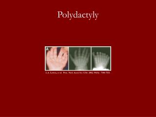
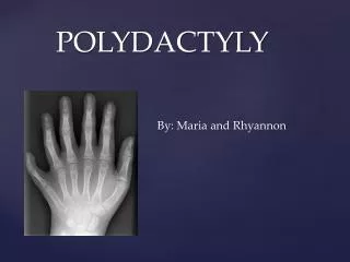
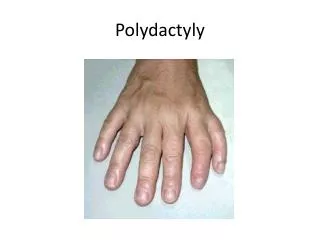
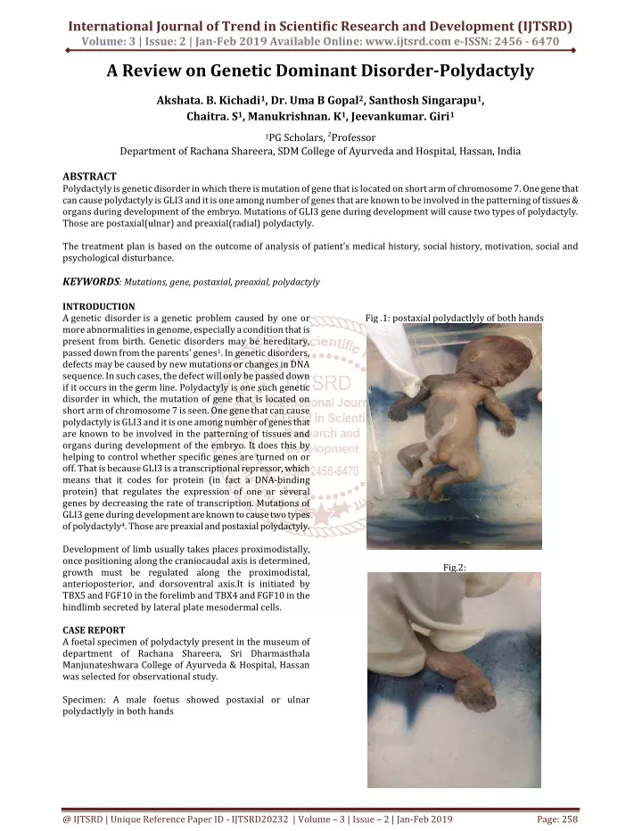
International Journal of Trend in Scientific Research and Development (IJTSRD) Volume: 3 | Issue: 2 | Jan-Feb 2019 Available Online: www.ijtsrd.com e-ISSN: 2456 - 6470 A Review on Genetic Dominant Disorder-Polydactyly Akshata. B. Kichadi1, Dr. Uma B Gopal2, Santhosh Singarapu1, Chaitra. S1, Manukrishnan. K1, Jeevankumar. Giri1 1PG Scholars,2Professor Department of Rachana Shareera, SDM College of Ayurveda and Hospital, Hassan, India ABSTRACT Polydactyly is genetic disorder in which there is mutation of gene that is located on short arm of chromosome 7. One gene that can cause polydactyly is GLI3 and it is one among number of genes that are known to be involved in the patterning of tissues & organs during development of the embryo. Mutations of GLI3 gene during development will cause two types of polydactyly. Those are postaxial(ulnar) and preaxial(radial) polydactyly. The treatment plan is based on the outcome of analysis of patient’s medical history, social history, motivation, social and psychological disturbance. KEYWORDS: Mutations, gene, postaxial, preaxial, polydactyly INTRODUCTION A genetic disorder is a genetic problem caused by one or more abnormalities in genome, especially a condition that is present from birth. Genetic disorders may be hereditary, passed down from the parents' genes1. In genetic disorders, defects may be caused by new mutations or changes in DNA sequence. In such cases, the defect will only be passed down if it occurs in the germ line. Polydactyly is one such genetic disorder in which, the mutation of gene that is located on short arm of chromosome 7 is seen. One gene that can cause polydactyly is GLI3 and it is one among number of genes that are known to be involved in the patterning of tissues and organs during development of the embryo. It does this by helping to control whether specific genes are turned on or off. That is because GLI3 is a transcriptional repressor, which means that it codes for protein (in fact a DNA-binding protein) that regulates the expression of one or several genes by decreasing the rate of transcription. Mutations of GLI3 gene during development are known to cause two types of polydactyly4. Those are preaxial and postaxial polydactyly. Development of limb usually takes places proximodistally, once positioning along the craniocaudal axis is determined, growth must be regulated along the proximodistal, anterioposterior, and dorsoventral axis.It is initiated by TBX5 and FGF10 in the forelimb and TBX4 and FGF10 in the hindlimb secreted by lateral plate mesodermal cells. CASE REPORT A foetal specimen of polydactyly present in the museum of department of Rachana Shareera, Sri Dharmasthala Manjunateshwara College of Ayurveda & Hospital, Hassan was selected for observational study. Specimen: A male foetus showed postaxial or ulnar polydactlyly in both hands Fig .1: postaxial polydactlyly of both hands Fig.2: @ IJTSRD | Unique Reference Paper ID - IJTSRD20232 | Volume – 3 | Issue – 2 | Jan-Feb 2019 Page: 258
International Journal of Trend in Scientific Research and Development (IJTSRD) @ www.ijtsrd.com eISSN: 2456-6470 Fig.3: There are three types of classification of polydactyly they are preaxial or radial,postaxial or ulnar and central. Among them preaxial is most common, refers to duplication of first digital rays.4 The foetal specimen found in museum of department of rachana shareer, SDMCA & H,Hassan it was found that the male foetus had postaxial polydactyly of both upper limbs. DISCUSSION Polydactyly follows an autosomal dominant pattern of inheritance which means that ?It is not sex linked, so that boys and girls may be equally affected and ?A child who has polydactyly parent has 50% chance of being polydactyly . Polydactyly is reported to occur in 1 in every 500 live births and hence it is not uncommon.5 Positioning of the limbs along the craniocaudal axis in the flank region of the embryo is regulated by the HOX gene expressed along this axis. These homeobox genes are expressed in overlapping patterns from head to tail with some having more cranial limits than others. For example, the cranial limit of expression of HOXB8 is at the cranial border of forelimbs, and misexpression of this gene alters the position of limbs. Once outgrowth is initiated, bone morphogenetic proteins(BMPs), expressed in ventral ectoderm, induce formation of apical endodermal ridge(AER) by signalling through the homeobox gene MSX2. Expression of radical fringe, in the dorsal half of the limb ectoderm, restricts the location of AER to the distal tip of the limbs. This gene induces expression of SER2, a homologue of drosophilia serrate, at the border between cells that are expressing radical fringe and those that are not. It is at this border that the AER is established. Formation of the border itself is assisted by expression of engrailed-1 in ventral ectoderm cells, because this gene represses expression of radical fringe. After the ridge is established, it expresses FGF4 and FGF8, which maintain the progress zone, the rapidly proliferating population of mesenchyme cells adjacent to the ridge. Distal growth of limb is then affected by this rapidly proliferating cells under the influence of the FGF’s. As growth occurs, mesenchymal cells at the proximal end of the progress zone become farther away from the ridge and its influence begin to slow down their division rates and differentiation. Patterning of the anteroposterior axis of the limb is regulated by the zone of polarizing activity(ZPA), a cluster of cells at the posterior border of the limb near the flank. These cells produce retinoic acid (vitamin A), which initiates expression of sonic hedgehog(SHH), a secreted factor that regulates the anteroposterior axis. Thus, for example, digits appear in the proper order, with the thumb on the radial(anterior) side. As the limb grows, the ZPA moves distal ward to remain in proximity to the posterior border of the AER. Misexpression of retinoic acid or SHH in the anterior margin of the limb containing a normally expressing ZPA in the posterior border results in mirror image duplication of limb structures. As per routine observation done in museum of department of Rachana Shareera ,SDMCA & H,Hassan it was found that the male foetus had postaxial polydactyly of both upper limbs. Polydactyly is the most common congenital anomaly of hand and foot. Embryologically Limb development begins with the activation of a group of a mesenchymal cells in the lateral plate mesoderm. It is said that a specialized gene called homeobox gene regulate the pattern in the formation of limb2. They start forming at the second month of intrauterine life. At the tip of each limb bud, the ectoderm is thickened to form the apical ectodermal ridge(AER). This ridge has an inducing effect on underlying mesenchyme causing it to remain undifferentiated and to proliferate, the progress zone. Areas away from the apical ridge undergo differentiation into cartilage, muscles, etc. In this manner ,development of the limb proceeds proximodistally.3 The forelimb bud appear a little earlier than the hindlimb buds. In 6week old embryos, the terminal portion of the limb buds becomes flattened to form the hand and footplates and is separated from the proximal segment by a circular constriction.6 As each forelimb bud grows, it becomes subdivided by constrictions into arm, forearm and hand. The hand itself soon shows outline of digits. The interdigital areas show cell death because of which the digits separate from each other. Similar changes occur in hindlimbs. While the limb buds are growing, the mesenchymal cells in the buds form cartilaginous models, which subsequently ossify to form the bones of the limbs. The limb buds are first directed forward and laterally from the body of the embryo. Each bud has preaxial border and postaxial border. The thumb and the great toe are formed on the preaxial border.3 During the seventh week of gestation, the limbs rotates in opposite directions. The upper limb rotates 90 degree laterally, so that the extensor muscles lie on the lateral and posterior surface, and the thumb lie laterally whereas the lower limb rotates approximately 90 degree medially, placing the extensor muscles on the anterior surface and the big toe medially.6Sometimes two AER are formed on the limb bud. This results in formation of super numerary limb3 @ IJTSRD | Unique Reference Paper ID - IJTSRD20232 | Volume – 3 | Issue – 2 | Jan-Feb 2019 Page: 259
International Journal of Trend in Scientific Research and Development (IJTSRD) @ www.ijtsrd.com eISSN: 2456-6470 The dorsoventral axis is regulated by BMP’s in the ventral ectoderm, Which induce expression of the transcription factor EN1. In turn, EN1 represses WNT7a expression, restricting it to the dorsal limb ectoderm. WNT7a is a secreted factor that induces expression of LMX1, a transcription factor containing a homeodomain, in the dorsal mesenchyme. LMX1 specifies cells to be dorsal, establishing the dorsoventral components. In addition, WTN7a maintains SHH expression in the ZPA and therefore indicatly affects anteroposterior patterning as well. These two genes are also intimately linked in signalling pathways in Drosophilia, and this interaction is conserved in vertebrates. In fact, all of the patterning genes in the limb have feedback loops. Thus, FGF’s in the AER activate SHH in the ZPA, while WNT7a maintains the SHH signal. Although patterning genes for the limbs axes have been determined, it is the HOX genes that regulate the types and shapes of the bones of the limb. Thus, HOX gene expression, which results from the combinatorial expression of SHH,FGFs, AND WNT7a, occurs in phases in three places in the limb that correspond to formation of the proximal(stylopod), middle(zeugopod), and distal(autopod) parts. Genes of the HOXA and HOXD clusters are the primary determinants in the limb, and variations in their combinatorial patterns of expression may account for differences in forelimb and hindlimb structures. Just as in the craniocaudal axis of the embryo, HOX genes are nested in overlapping patterns of expression that somehow regulate patterning. Factors determining forelimb versus hindlimb are the transcription factors TBX5(forelimb) and TBX4 together with PITX1(hindlimbs)6. CONCLUSION Polydactyly is the most common congenital anomaly of the hand and foot. The frequency of polydactyly varies widely among populations. It may be an isolated condition or part of a congenital syndrome. Polydactyly is generally classified into three major groups: medial ray (preaxial), central ray and lateral ray(postaxial). The deformity is cosmetically unacceptable in many cultures and this in turn leads to psychological burden. Polydactyly often causes social embarrassment and results in compromised to move the hand independently. It often has severe impact on patients self-esteem and hampers his or her quality of life. Hence the treatment plan is formulated and tailored specifically for each individual patient. Though evaluation and diagnosis are among most important aspects of overall patient’s management, the treatment plan is based on the outcome of analysis of patient’s medical history, social history, motivation, social and psychological disturbances. A patients ultimate satisfaction with treatment outcome often depends upon attention to the patients’ chief concerns7. In the dead foetal specimen mounted in museum of SDMCA & H, Hassan, it was observed that the polydactyly was of post axial or ulnar polydactyly in both upper limbs. Radiographs of the affected limb are recommended to show whether the rudimentary dgit contains skeletal elements. The degree of deviation of the digit and the size of the articulating metacarpal or metatarsal also may be helpful and surgical planning. REFERENCES [1]https://en.m.wikipedia.org/wiki/Genetic_disorder. [2]The developing human,clinically oriented embryology ,moore & persaud, 7th edition,pg-410. [3]Human edition,pg-122 embryology,inderbir singh,G.P Pal,8th [4]Left handed polydactyly: a case report, Nicola mumali et all [5]What causes polydactyl written by paul Arnold.edited by paul Arnold.updated:8/29/2009,bright https://www.brighthub.com/science/genetics/articles /47390.aspx hub. [6]Langman’s Medical embryology, T.W.Sadler,eleventh edition,south Asian edition,published by wolters Kluwer(india) pvt.ltd.,new delhi.chapter 9,pg 134-141 [7]Left handed polydactyly: a case report, Nicola mumali et all. @ IJTSRD | Unique Reference Paper ID - IJTSRD20232 | Volume – 3 | Issue – 2 | Jan-Feb 2019 Page: 260