Download
1 / 9
90 likes | 100 Vues
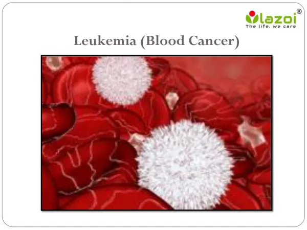
Leukemia is a gathering of malignancy began from bone marrow and will deliver impact on white platelet which will thus lead to anomaly. Presently the development of impact will continue by the undeveloped and juvenile WBC. Leukemia is an expansive term covering a range of infections. The focal point of enthusiasm for oncology especially in leukemia vitamin C has the best technique by which it demonstrates the more noteworthy outcome. An undeveloped cell takes amazingly elevated amounts of Ascorbic acid, which at that point allot their capacity and subdue the improvement of leukemia. Vitamin C levels manage HSC numbers and capacity. At the point when ascorbate levels are reduced, it can give the power to the deactivation of a catalyst called Tet2. The inactivation of TET2 is represented by change and insufficiency of vitamin C may constrain Tet2 work in tissues in a way that builds the danger of leukemia. TET2 function is actually strengthen by ascorbate level. By encouraging DNA demethylation, tremendous dose of ascorbate treatment can persuade stem cells to develop and the growth of cancerous stem cells will retarded by the tremendous dose of ascorbate level as investigated in mice. Tremendous dose of ascorbate might become a safe treatment for blood diseases caused by TET2 deficient leukemia stem cells, most likely in combination with other targeted therapies. Tarun Gupta | Virendra Kumar Vishwakarma | Manoj Kumar Maurya | Khushdil Khan "Anti-cancer Activity of Vitamin-C Against Leukemia: A Review" Published in International Journal of Trend in Scientific Research and Development (ijtsrd), ISSN: 2456-6470, Volume-2 | Issue-4 , June 2018, URL: https://www.ijtsrd.com/papers/ijtsrd14250.pdf Paper URL: http://www.ijtsrd.com/biological-science/biotechnology/14250/anti-cancer-activity-of-vitamin-c-against-leukemia-a-review/tarun-gupta<br>

E N D
International Research Research and Development (IJTSRD) International Open Access Journal International Open Access Journal International Journal of Trend in Scientific Scientific (IJTSRD) ISSN No: 2456 - 6470 | www.ijtsrd.com | Volume C Against Leukemia: A Review ISSN No: 2456 www.ijtsrd.com | Volume - 2 | Issue – 4 Anti-cancer Activity of Vitamin cancer Activity of Vitamin-C Against Leukemia 1Tarun Gupta, 2Virendra Kumar Vishwakarma, 1,4Department of Biotechnology 2,3Department of Advanced Science Biotechnology, NIET, Nims Unive Department of Advanced Science Biotechnology, NIET, Nims University Rajasthan, Jaipur Virendra Kumar Vishwakarma, 3Manoj Kumar Maurya Department of Biotechnology, Mohanlal Sukhadia University, Udaipur, Rajasthan Manoj Kumar Maurya, 4Khushdil Khan , Rajasthan, India rsity Rajasthan, Jaipur, Rajasthan, India cancer, Bladder cancer, Bone cancer, Blood cancer, Breast cancer, Colon cancer, Ovarian cancer, Liver cancer, Leukemia, Thyroid cancer, Prostate cancer and so on. Cancer hurts the body when adjusted cells separate wildly to shape bumps or masses of tissue called tumors (with the exception of on account of leukemia where cancer disallows ordinary blood work by anomalous cell division in the circulation system) . At the point when a tumor viably spreads to different parts of the body and develops, attacking and pulverizing other solid tissues, it is said to have metastasized. Leukaemia, leukemia, is a gathering of tumors that typically start in the bone marrow and result in high quantities of irregular white platelets. This procedure itself is called metastasis and the outcome is a genuine condition that is extremely hard to treat and intense or incessan ailment in people portrayed by an anomalous increment in the quantity of white platelets in tissues and regularly in the blood leukemia is unknown. A combination of factors and environmental (non believed to play include smoking, ionizing radiation (such as benzene), prior chemotherapy, and syndrome [1]. Stem cells are undifferentiated natural cells that can separate into particular cells and can partition (through mitosis) to deliver more stem cells. 2. Vitamin C The substrates, intermediates and products of cellular metabolism have the potential to in identity and transformation to cancer C, otherwise called ascorbic acid and L is a vitamin found in sustenance and utilized as a dietary supplement. Vitamin C is a basic supplement for specific creatures including people. The term for specific creatures including people. The term ABSTRACT Leukemia is a gathering of malignancy began from bone marrow and will deliver impact on white platelet which will thus lead to anomaly. Presently t development of impact will continue by the undeveloped and juvenile WBC. Leukemia is an expansive term covering a range of infections. The focal point of enthusiasm for oncology leukemia vitamin C has the best technique by which it demonstrates the more noteworthy outcome. undeveloped cell takes amazingly elevated amounts of Ascorbic acid, which at that point allot their capacity and subdue the improvement of leukemia. Vitamin C levels manage HSC numbers and capacity. At the point when ascorbate levels are reduced, it can give the power to the deactivation of a catalyst called Tet2. The inactivation of TET2 is represented by change and insufficiency of vitamin-C may constrain Tet2 work in tissues in a way that builds the danger of leukemia. TET2 function is actually strengthen by ascorbate level. By encouraging DNA demethylation, tremendous-dose of ascorbate treatment can persuade stem cells to develop and the growth of cancerous stem cells will retarded by the tremendous dose of ascorbate level as investigated in mice. Tremendous dose of ascorbate might become a safe treatment for blood diseases caused by TET2-deficient leukemia stem cells, most likely in combination with other targeted therapies. Keywords - Leukemia, Blast cells, Vitamin DNA Methylation is a gathering of malignancy began from will deliver impact on white platelet anomaly. Presently the cancer, Bladder cancer, Bone cancer, Blood cancer, Breast cancer, Colon cancer, Ovarian cancer, Liver a, Thyroid cancer, Prostate cancer and so on. Cancer hurts the body when adjusted cells separate wildly to shape bumps or masses of tissue called tumors (with the exception of on account of leukemia where cancer disallows ordinary blood work ell division in the circulation system) . At the point when a tumor viably spreads to different parts of the body and develops, attacking and pulverizing other solid tissues, it is said to have metastasized. Leukaemia, athering of tumors that typically start in the bone marrow and result in high quantities of This procedure itself is called metastasis and the outcome is a genuine condition that is extremely hard to treat and intense or incessant ailment in people portrayed by an anomalous increment in the quantity of white platelets in tissues and regularly in the blood. The exact cause of A combination of genetic and environmental (non-inherited) factors are believed to play ionizing radiation, some chemicals ), prior chemotherapy, and Down Stem cells are undifferentiated natural cells that can separate into particular cells and can partition (through mitosis) to deliver more stem cells. continue by the undeveloped and juvenile WBC. Leukemia is an range of infections. The focal point of enthusiasm for oncology especially in has the best technique by which it demonstrates the more noteworthy outcome. An amazingly elevated amounts of at point allot their capacity and subdue the improvement of leukemia. Vitamin C levels manage HSC numbers and capacity. At the point when ascorbate levels are reduced, it can give the power to the deactivation of a catalyst called Tet2. TET2 is represented by change additionally additionally spelled spelled C may constrain Tet2 work in tissues in a way that builds the danger of TET2 function is actually strengthen by ascorbate level. By encouraging DNA demethylation, rbate treatment can persuade stem cells to develop and the growth of cancerous stem cells will retarded by the tremendous dose of ascorbate level as investigated in mice. Tremendous dose of ascorbate might become a safe treatment for a a role. role. Risk factors deficient leukemia stem cells, most likely in combination with other Leukemia, Blast cells, Vitamin-C, Tet2, he substrates, intermediates and products of cellular metabolism have the potential to influence cellular identity and transformation to cancer [2-3]. Vitamin C, otherwise called ascorbic acid and L-ascorbic acid, is a vitamin found in sustenance and utilized as a Vitamin C is a basic supplement 1. Introduction Cancer is a class of disease described by uncontrolled cell development.There are more than 100 distinct sorts of cancer, and each is characterized by the kind of cell that is fundamentally overstated like Anal of cell that is fundamentally overstated like Anal Cancer is a class of disease described by uncontrolled There are more than 100 distinct sorts of cancer, and each is characterized by the kind @ IJTSRD | Available Online @ www.ijtsrd.com @ IJTSRD | Available Online @ www.ijtsrd.com | Volume – 2 | Issue – 4 | May-Jun 2018 Jun 2018 Page: 1264
International Journal of Trend in Scientific Research and Development (IJTSRD) ISSN: 2456-6470 vitamin C envelops a few vitamers that have vitamin C action in creatures. Ascorbate salts, for example, sodium ascorbate and calcium ascorbate are utilized as a part of some dietary supplements. These releases ascorbate upon retention. Ascorbate and ascorbic acid are both ordinarily display in the body, since the structures interconvert according to pH. Oxidized types of the particle, for example, dehydroascorbic acid are changed over back to ascorbic acid by decreasing operators [4]. Vitamin C can be produced by most animals and plants from D-glucose and D- galactose, whereas it is not produced in humans due to the lack of L-gluconolactone oxidaseenzyme; and therefore, it should be taken externally [5]. The biological importance of vitamin C is that it assumes a cofactor part, as a reducing specialist, in different enzymatic reactions. Human cells can't combine Vit C, and accordingly, it should constantly be re- established through the eating. Extreme vitamin C deficiency prompts gum recession, shortcoming, laziness, and scurvy characterized by simple draining and wounding. The symptoms of scurvy develop as a result of weakened connective tissue in the body, and vitamin C is given to patients with scurvy for strengthening their connective tissues [6]. Vit C has emerged as a key regulator of stem cell identity/behavior, influencing pluripotency, self- renewal, and differentiation. The World Health Organization (WHO) recommends a daily intake of 45 mg vit C per day for healthy adults [7]. 3. Origin of Vitamin C After 1753 citrus organic products were started to avert scurvy; and upon that, the method for the way to the disclosure of vitamin C was opened. In 1912, the researcher Casimir Funk instituted the expression "antiscorbutic vitamin" to allude this compound contained in products of the soil. In 1920, the British organic chemist Jack Drummond utilized the expression "vitamin C" to allude this compound [8-9]. Vitamin C was discovered in 1912, isolated in 1928 and synthesized in 1933, making it the first vitamin to be synthesized [10]. Ascorbic acid was first isolated from natural sources and structurally characterized by Szent-Gyorgyi, Waugh and King [11-12]. Few warm blooded animals are helpless to scurvy as their liver does not produce the catalyst L-gulonolactone oxidase, the remainder of the chain of four compounds that blend vitamin C. High-dose vitamin C started to be attempted by the Scottish specialist Ewan Cameron without precedent for 1970, in his examinations on growth patients. 4. Risk Factors for Leukemia The cancerous state of leukemia initiates when the DNA of one or more white blood cells is damaged or mutated. In many instances, DNA damage results in the activation of oncogenes (cancer-promoting genes) and deactivation of tumor suppressor genes (cancer- preventing genes). Age: About 60-70% of leukemics are older than 50 Previous cancer treatment: Individuals who've had certain sorts of chemotherapy and radiation treatment for different tumors have an expanded danger of building up specific kinds of leukemia. Genetic disorders: Genetic abnormalities seem to play a role in the development of leukemia. Certain genetic disorders, such as Down syndrome, are associated with an increased risk of leukemia. Smoking: It is reported that many of the adult cases with leukemia are cigarette smokers. Exposure to certain chemicals: Exposure to certain chemicals, such as benzene — which is found in gasoline and is used by the chemical industry — is linked to an increased risk of some kinds of leukemia 5. Symptoms Fig.1 - Symptoms of Leukemia Leukemia symptoms vary, depending on the type of leukemia. Common leukemia signs and symptoms include fig.1. @ IJTSRD | Available Online @ www.ijtsrd.com | Volume – 2 | Issue – 4 | May-Jun 2018 Page: 1265
International Journal of Trend in Scientific Research and Development (IJTSRD) ISSN: 2456-6470 -Fever or chills Myeloid leukemia: Leukemias that start in early forms of myeloid cells – the cells that make white blood cells (other than lymphocytes), red blood cells, or platelet-making cells (megakaryocytes) – are myeloid leukemias (also known as myelocytic, myelogenous, or non-lymphocytic leukemias). Lymphocytic leukemia: Leukemias that start in immature forms of lymphocytes lymphocytic leukemias (also known as lymphoid or lymphoblastic leukemias). [13]. Specific types Acute lymphoblastic leukemia (ALL) is the most common type of leukemia in young children. It also affects adults, especially those 65 and older. Standard treatments involve chemotherapy and radiotherapy. The survival rates vary by age: 85% in children and 50% in adults.[14] Subtypes include precursor B acute lymphoblastic leukemia, precursor lymphoblastic leukemia, Burkitt's leukemia, and acute biphenotypic leukemia. Chronic lymphocytic leukemia (CLL) most often affects adults over the age of 55. It sometimes occurs in younger adults, but it almost never affects children. Two-thirds of affected people are men. The five-year survival rate is 75%.[15] It is incurable, but there are many effective treatments. One subtype is B-cell prolymphocytic leukemia, a more aggressive disease. Acute myelogenous leukemia (AML) occurs more commonly in adults than in children, and more commonly in men than women. It is treated with chemotherapy. The five-year survival rate is 40%, except for APL (Acute Promyelocytic Leukemia), which has a survival rate greater than 90%. Subtypes of AML include acute promyelocytic leukemia, acute myeloblastic leukemia, and acute megakaryoblastic leukemia. Chronic myelogenous mainly in adults; a very small number of children also develop this disease. with imatinib (Gleevec in United States, Glivec in Europe) or other drugs. The five-year survival rate is 90%. Myeloblast Disorders Blast percentage plays a central role in the diagnosis and classification of myelodysplastic syndromes (MDS). In the case of acute myelogenous (AML) and myelodysplastic syndromes (MDS), there is an overproduction of abnormal myeloblasts. These - Persistent fatigue, weakness -Frequent or severe infections -Swollen lymph nodes -enlarged liver or spleen -Easy bleeding. are called -Recurrent nosebleeds -Tiny red spots in your skin (petechiae) -Excessive sweating, especially at night -Bone pain or tenderness 6. Different Classes of Leukemia Types of leukemia are grouped by the type of cell affected and by the rate of cell growth. Leukemia can be either acute or chronic. Acute leukemia involves an overgrowth of very immature blood cells. This condition is life threatening because there are not enough mature blood cells to prevent anemia, infection and bleeding. A diagnosis of acute leukemia is made when there are 20% or more blasts or immature cells in the bone marrow. A.Acute leukemia versus chronic leukemia- The first factor in classifying leukemia is whether most of the abnormal cells are mature (look like normal white blood cells) or immature (look more like stem cells). Acute leukemia: In acute leukemia, the bone marrow cells cannot mature properly. Immature leukemia cells continue to reproduce and build up. Without treatment, most people with acute leukemia would live only a few months. [13] Chronic leukemia: In chronic leukemia, the cells can mature partly but not completely. These cells may look fairly normal, but they generally do not fight infection as well as normal white blood cells do. They also live longer, build up, and crowd out normal cells. [13] B.Myeloid leukemia leukemia- The second factor in classifying leukemia is the type of bone marrow cells that are affected T acute leukemia (CML) occurs It is treated versus lymphocytic acute leukemias and leukemia @ IJTSRD | Available Online @ www.ijtsrd.com | Volume – 2 | Issue – 4 | May-Jun 2018 Page: 1266
International Journal of Trend in Scientific Research and Development (IJTSRD) ISSN: 2456-6470 cells are unable to develop further into mature white blood cells. Acute myeloid leukemia is a type of cancer that goes by several other names, such as acute myelocytic leukemia, acute myelogenous leukemia, acute granulocytic leukemia, acute non-lymphocytic leukemia, or sometimes just AML. It is most common in older people. Most cases of AML develop from cells that would turn into white blood cells other than lymphocytes, however, some cases of AML develop in other types of blood-forming cells [16]. who deficiency; and the risk of renal failure in those who have renal disorders [19-20]. Vitamin C acidifies the urine, it can cause the formation of kidney stones; and on the other hand, a case of hyperoxaluria may emerge due to oxalic acid formed as a result of vitamin C metabolism [21]. In a study published in 2000, the tolerable upper limit of vitamin C was determined for the first time to be 2 g, and amounts exceeding this limit were reported to have a laxative effect. Diarrhoea is already the most common side effect of high doses of vitamin C [22]. Tremendous dose of vitamin C in combination with the chemotherapy will produce a danger effect and causes toxicity. There are studies showing that the use of high dose vitamin C in combination with certain chemotherapy drugs increases the toxicity. It has been shown that the use of high-dose vitamin C in combination with arsenic trioxide in acute myeloid leukemia, refractory metastatic colon cancer, and metastatic melanoma causes serious side effects and leads to the disease's progression [23-24] Inflammation and Cancer. Leukocytes and other phagocytic cells combat bacteria, parasites, and virus infected cells by destroying them with nitrogen oxide and superoxide, which react to form peroxynitrite, a powerful mutagenic oxidizing and nitrating agent; hypochlorite, a mutagenic chlorinating and oxidizing agent; and hydrogen peroxide, a mutagenic oxidizing agent. These oxidants protect humans from immediate death from infection but also cause oxidative damage to DNA, mutation, and chronic cell killing with compensatory cell division, thereby contributing to the carcinogenic process. Antioxidants appear to inhibit some of the pathology of chronic inflammation [25] fig.2. have Glucose-6-phosphate dehydrogenase Myelodysplastic syndrome is a group of disorders that affects the production of new blood cells in the bone marrow. In these diseases, the bone marrow produces abnormal blast cells that fail to mature properly and are unable to function. These abnormal blasts begin to take over the bone marrow and prevent the production of adequate numbers of other types of blood cells, such as platelets, red blood cells, and healthy white blood cells. In fact, production of leukemic blasts may get so out of hand that the immature cells spill out from the bone marrow into the circulation. The presence of blast cells on a complete blood count (CBC) is therefore very suspicious for leukemia. Blast cells are not typically found in the circulating blood of healthy individuals [17]. 7. Effect of low dose and high dose of ascorbate The low concentration of vitamin c will produce a tremendous effect on the body by the deficiency of the vitamin c scurvy will produced. The amount <45 mg C will cause a great effect and promotes the class of disease for attacking the body .As said that vitamin C is water soluble in nature so the no stored form of vitamin c is found usually in body .It has been identified that low concentration of vitamin c will cause spongy and sore gums, loose teeth. By the effect of low dose the fragile blood vessels are formed and finally cause the anaemia that will lead to the death in severe conditions. The tumours will form and will cause haemorrhage. Most of these above mentioned effects are related to impairment in synthesis of collagen and/or the antioxidant property of vitamin C. Although intake of high doses of vitamin C is alleged to be harmless, based on that it is a water soluble vitamin and is not stored in the body, there are many side effects and drug interactions reported in the literature [18], It has been reported that high-dose vitamin C could increase haemolytic anaemia in those Fig. 2 Inflammation and cancer @ IJTSRD | Available Online @ www.ijtsrd.com | Volume – 2 | Issue – 4 | May-Jun 2018 Page: 1267
International Journal of Trend in Scientific Research and Development (IJTSRD) ISSN: 2456 International Journal of Trend in Scientific Research and Development (IJTSRD) ISSN: 2456 International Journal of Trend in Scientific Research and Development (IJTSRD) ISSN: 2456-6470 Tissue events Chronic lymphocytic leukemia cells circulating in the peripheral blood are the tip of the iceberg. The most significant pathophysiological events occur in tissues [25] where leukemic cells: (i) are activated by exposure to antigens, although it is still unclear where and how this exposure takes place. It is also unclear how B-cell antigen receptor (BCR) stimulation may translate into either cell proliferation or cell anergy and how these processes are mediated by various signal transduction pathways; (ii) receive the proper T-cell help, if and when needed, to be selected for clonal expansion; (iii) proliferate in specific niches, the pseudo follicular proliferation centers, which are not detected in any other B malignancy, but are observed in inflamed tissues of patients with systemic autoimmune/inflammatory disorders; and (iv) interact with stromal cells that favor cell accumulation. CLL cells not only interact with but also shape microenvironments by recruiting activated T cells and stromal cells by means of chemokines and chemokine receptors. [26-27] 8. Vitamin C- A Potent Antitumor Agent Ascorbate was originally considered as an adjuvant with favorable biological properties. Cameron and Pauling suggested ascorbate increased extracellular collagen production and strengthened the extracellular matrix, thus walling in tumors [28]. Proposed mechanisms of vitamin C activity in the prevention and treatment of cancer include: (1) enhancement of the immune system by increased lymphocyte production; (2) stimulation of collagen formation, necessary for “walling off” tumors; (3) inhibition of hyaluronidase, keeping the ground substance around the tumor intact and preventing metastasis [29] (4) inhibition of oncogenic viruses; (5) correction of an ascorbate deficiency, cancer patients (6) expedition of wound healing after cancer surgery [30] (7) enhancement of the effect of certain chemotherapy drugs. H2O2 plays a vital role as a cytotoxic mediator high-dose vitamin C therapy fig.3. In vitro mediated by H2O2 rather than ascorbic radicals and H2O2 formation results in selective cytotoxicity. The H2O2 formed from pharmacological ascorbate concentrations diffuses into cells.[31]. within the cells may cause breaks in DNA and mitochondria and the mitochondria in some cancer cells may have increased sensitivity to H cells may have increased sensitivity to H2O2 [32] cells circulating in blood are the tip of the iceberg. The most significant pathophysiological events occur in (i) are activated by exposure to antigens, although it is still unclear where and how this exposure takes place. cell antigen receptor (BCR) stimulation may translate into either cell proliferation or cell anergy and how these processes are mediated by various signal transduction pathways; (ii) receive cell help, if and when needed, to be for clonal expansion; (iii) proliferate in specific niches, the pseudo follicular proliferation centers, which are not detected in any other B-cell malignancy, but are observed in inflamed tissues of patients with systemic autoimmune/inflammatory s; and (iv) interact with stromal cells that favor cell accumulation. CLL cells not only interact with but also shape microenvironments by recruiting activated T cells and stromal cells by means of chemokines and chemokine Fig.3 - Antitumor activity of ascorbate [33] Antitumor activity of ascorbate [33] their their conducive conducive 9. TET2 gene and its role TET2 is implicated in a variety of hematopoietic malignancies, particularly myeloid malignancies. Ascorbate is a cofactor for several dependent dioxygenases15, [34], a cofactor for prolyl hydroxylases that degrade hypoxia inducible factors (HIFs) [35], but HIF HIF-2α are not required for HSC function Despite our inability to detect an effect of ascorbate depletion on the function of these enzymes, they could potentially contribute to the effects of ascorbate depletion on HSCs. Ascorbate promotes the activity of Tet enzymes in culture [37- unclear whether ascorbate limits Tet function Tet enzymes convert 5-methylcytosine (5mC) to 5 hydroxymethylcytosine (5hmC), and subsequently catalyse some of the steps towards un cytosine [39-40] fig. 4. Mutations that inactivate Tet2 leukaemogenesis26 that increase HSC frequency and self-renewal [41-43] and cause leukaemia when combined with other mutations 5hmC exhaustion in an assortment of human malignancies recommends diminished TET action in tumor cells. The expansion of vitamin C to ES cells advances DNA demethylation through expanded TET activity. is implicated in a variety of hematopoietic malignancies, particularly myeloid malignancies. Ascorbate is a cofactor for several α -ketoglutarate- A Potent Antitumor Agent Ascorbate was originally considered as an adjuvant with favorable biological properties. Cameron and Pauling suggested that [34], Ascorbate can act as response response modifying modifying a cofactor for prolyl hydroxylases that degrade nducible factors (HIFs) [35], but HIF-1α and are not required for HSC function [36]. Despite our inability to detect an effect of ascorbate depletion on the function of these enzymes, they could potentially contribute to the effects of ascorbate letion on HSCs. Ascorbate promotes the activity collagen production and strengthened the extracellular matrix, thus walling Proposed mechanisms of vitamin C activity in the prevention and treatment of cancer include: (1) enhancement of the immune system by increased n; (2) stimulation of collagen formation, necessary for “walling off” tumors; (3) inhibition of hyaluronidase, keeping the ground substance around the tumor intact and preventing [29] (4) inhibition of oncogenic viruses; (5) -38], although it remains unclear whether ascorbate limits Tet function in vivo. methylcytosine (5mC) to 5 - hydroxymethylcytosine (5hmC), and subsequently talyse some of the steps towards un-methylated often seen in nd healing after Tet2 are early events in [30] (7) enhancement of the effect of leukaemogenesis26 that increase HSC frequency and 43] and cause leukaemia when combined with other mutations [44-45]. Likewise, 5hmC exhaustion in an assortment of human malignancies recommends diminished TET action in . The expansion of vitamin C to ES cells advances DNA demethylation through expanded TET plays a vital role as a cytotoxic mediator in vitro, killing is rather than ascorbic radicals and formation results in selective cytotoxicity. The formed from pharmacological ascorbate concentrations diffuses into cells.[31]. The H2O2 within the cells may cause breaks in DNA and mitochondria and the mitochondria in some cancer @ IJTSRD | Available Online @ www.ijtsrd.com @ IJTSRD | Available Online @ www.ijtsrd.com | Volume – 2 | Issue – 4 | May-Jun 2018 Jun 2018 Page: 1268
International Journal of Trend in Scientific Research and Development (IJTSRD) ISSN: 2456-6470 The role of DNA methylation as an epigenetic factor has been proven in regulating genes involved in tumor genesis; such as breast, prostate and colorectal cancer, as well as in hematologic malignancies including MDS (Myelodysplastic Syndromes), AML (Acute Myelogenous Leukemia), Myelogenous Leukemia), Nevertheless, effective mechanisms related to DNA methylation changes are yet to be discovered [57]. Also, through multiple histone modifications, histone methylation has been considered as an active element regulating transcriptional activity of genes [58]. DNA methylation of cytosine C5 position is one of the key epigenetic alterations in CpG dinucleotides and plays the main role in normal development as well as tumor genesis [59]. Methyl groups add to DNA through certain enzymes known as DNA. These enzymes are responsible for the stability of methylation patterns during development (DNMT3A, DNMT3B and their co-factor DNMT3- like); and maintenance of methylation during replication (DNMT1) [60]. The majority of CpG sites were shown to be methylated in mammalian cells. However, there is evidence showing that CpG islands have been mostly deviated from the genomic regular pattern, creating CpG rich regions which are often un methylated [57]. 11. Vitamin-C as a multi-targeting anti-cancer agent High-dose intravenous ascorbate (IVC) has attracted increasing interests as a low-toxic cancer therapy. IVC bypasses bioavailability barriers of oral ingestion, provides pharmacologic concentrations in tissues, and exhibits selective cytotoxic effects in cancer cells through peroxide formation. The selectivity is related to the mechanisms of action. It is postulated that ascorbate-induced Reactive Oxygen Species (ROS) has multiple mechanisms of action that preferably influence cancer cells- (1) Ascorbate-produced ROS incites DNA harm. Downstream to DNA harm, cell NAD+ diminishes as an impact of poly ADP ribose polymerase (PARP) activation. Reduction glyceraldehyde 3-phosphate (GAPDH) movement and exhausts ATP in tumor cells, while ordinary cells keep up their ATP levels. This marvel has its root in dysregulated glucose digestion in disease cells, known as the Warburg 10. Regulators of TET activity While different cell types seem to express different amounts of each TET enzyme, it has become clear that post-translational regulation is critical in controlling TET activity and targeting to genetic loci. All TET enzymes contain one Cys-rich domain and two double-stranded β-helix (DSBH) domains that display the core catalytic domains, which act in a Fe(II) and 2-oxoglutarate (2-OG, also called as α- ketoglutarate) dioxygenases-dependent manner [46]. Mono ubiquitinylation at a conserved lysine residue (residue 1299 in TET2) [47-48] or binding of ascorbic acid in this catalytic domain directly facilitates TET catalytic activity by stabilizing Fe(II) association with the enzyme [49-50]. An important regulatory control on TET activity has been recently discovered with the finding of Isocitrate Dehydrogenase-1and -2 (IDH1/2) mutations in a variety of tumors, including AML and MDS. IDH1/2 are enzymes that play an important role in the tricarboxylic acid cycle (TCA). Heterozygous, gain of function mutations in these enzymes have been found in high frequency in myeloid malignancies [51-52]. These mutations cause IDH1/2 to produce an oncometabolite, 2-hydroxyglutarate (2-HG), instead of 2-OG [53]. This is a competitive antagonist of 2- OGT enzymes, which in turn leads to severely reduced TET enzyme activity. It is found that IDH1/2 mutations in AML patients lead to genome wide DNA hyper methylation signatures [54]. While the population of patients with genetic IDH1/2 mutations does not overlap with TET2 mutations, these different mutations have essentially synonymous hyper methylation signatures. This suggests that IDH1/2 and TET2 mutations phenocopy one another and therefore do not confer additional selective advantages during clonal evolution in these diseases. CML and (Chronic [56]. ALL of NAD+ hindered dehydrogenase Fig.4 Mechanism of TET2 gene to activate the vitamin C activity [55] @ IJTSRD | Available Online @ www.ijtsrd.com | Volume – 2 | Issue – 4 | May-Jun 2018 Page: 1269
International Journal of Trend in Scientific Research and Development (IJTSRD) ISSN: 2456-6470 Effect, whereby malignancy cells rely upon a bigger extent on glycolysis for ATP, though typical cells depend more on oxidative phosphorylation. (2) Absence of NAD+ represses movement of Sirt- 2(sirtuin-2), a tubulin deacetylase, and subsequently increments tubulin acetylation, which thus upsets progression of microtubules. This impacts tumor cells that are currently experiencing mitosis and movement. (3) At the point when PARP (Poly [ADP-ribose] polymerase ) is hindered, excessive DNA harm results in cell death. Further, ascorbate inhibited Epithelial-Mesenchymal change (EMT), a critical procedure adding to growth metastasis. At last, ascorbate improved collagen synthesis in tumor stroma. In spite of the dubious reports on the impact of raised collagen in tumor movement, the expanded collagen by ascorbate treatment is related with limitation of tumor invasion in our creature analyze and in patient. Taken together, these data show multi- targeting effects of death/inhibition in cancer cells relative to normal cells. With minimal toxicity, the multi-targeting mechanism of ascorbate is advantageous because it could decrease the likelihood of resistance, and provides multiple opportunities for combination with standard chemo and radiation therapies [61]. References: 1)"Leukemia". NCI. Archived from the original on 27 May 2014. Supplements.2nd ed. New York, NY: Informa Healthcare; pp 821-831. 7)Vitamin and Mineral Requirements in Human Nutrition , World Health Organization. 2nd ed. 2004, 138. 8)J. Lind and A. Millar. A Treatise of the Scurvy. London. 1753:149. 9)L. Rosenfeld. 1997. Vitamin-vitamin. The early years of discovery. Clin Chem.vol 43(4) pp 680- 685. 10)V.R. Agriculture, Forestry and Fisheries in Human Nutrition - Volume IV. EOLSS Publications. p. 121. Squires. 2011. The Role of Food, 11)J.L. Svirbely and G. Szent. 1932. The chemical nature of vitamin C. Biochem J. vol 26 pp 865- 870. 12)W.A. Waugh, and C.G. King.1932.Isolation and identification of vitamin C. J Biol .Chem. vol 97, pp 325-331. ascorbate that favor 13)A.S Advani, S. McDonough, S. Coutre et al. 2014 .SWOG S0910: A phase 2 trial of clofarabine/cytarabine/epratuzumab relapsed/refractory leukaemia. Br J Haematol. vol 165(4), pp 504- 509. for acute lymphocytic 14)Finding Cancer Statistics » Cancer Stat Fact Sheets »Chronic Lymphocytic Archived 16 April 2008 at the Wayback Machine. National Cancer Institute. Leukemia 2)L. Dang et al. 2009. Cancer-associated IDH1 mutations produce 2-hydroxyglutarate. Nature vol. 462, pp. 739–744. 15)G.A. Colvin and G.J. Elfenbein. 2003. "The latest treatment advances for acute myelogenous leukemia". Medicine and Health, Rhode Island. vol. 86 (8) , pp 243–6. 3)M.M. Mihaylova,. D. M Sabatini and O.H. Yilmaz, 2014. Dietary and Metabolic Control of Stem Cell Function in Physiology and Cancer Cell. Stem Cell vol.14(3), pp 292–305. 16)R.A. Larson and M.M. Le Beau.2011. Prognosis and Therapy When Acute Promyelocytic Leukemia and Other “Good Risk” Acute Myeloid Leukemias Occur as a Therapy-Related Neoplasm. Mediterranean. Journal of Hematology and Infectious Diseases. vol 3(1), pp 1-7. 4)Dietary Reference Intakes for Vitamin C, Vitamin E, Selenium, and Carotenoids. Institute of Medicine 2000.Washington, D.C.: The National Academies Press. Pp.95–185. Myeloid 17)M.F. Zahid, A. Parnes, B.N. Savani, M.R. Litzow and S.K. Hashmi.2016. Therapy-related myeloid neoplasms - what have we learned so far? World Journal of Stem Cells.vol 8(8) pp 231-242. 5)K.A. Naidu. 2003. Vitamin C in human health and disease is still a mystery? An overview. Nutr. J, vol. 2(7), pp 1-10 6)S. Padayatty, M.G. Espey and M. Levine. 2010. Vitamin C, Encyclopedia 18)Ahmet Unlu, M.D. Onder Kirca, M.D, Mustafa Ozdogan, M.D and Erdinç Nayır, M.D. 2016. of Dietary @ IJTSRD | Available Online @ www.ijtsrd.com | Volume – 2 | Issue – 4 | May-Jun 2018 Page: 1270
International Journal of Trend in Scientific Research and Development (IJTSRD) ISSN: 2456-6470 29)E. Cameron and L. Pauling.1973. Ascorbic acid and the glycosaminoglycans. Oncology. vol.27, pp 181-192. High-dose vitamin C and cancer. Journal of Oncological Science . vol (1), pp 10-12. 19)S.J. Padayatty et al. 2010. Intravenous use by complementary and practitioners and adverse effects. PLOS One.vol.5 (7), pp 1-8. 30)V.N. Sukolinskii and T.S. Morozkina.1989. Prevention of postoperative complication in patients with stomach cancer using an antioxidant complex.Vopr Onkol. vol. 35, pp 1242-1245. alternative medicine 20)D.C. Rees, H. Kelsey and J.D. Richards. 1993. Acute haemolysis induced by high dose ascorbic acid in glucose-6-phosphate deficiency. British Medical Journal. vol 306, pp 841-842. 31)F. Antunes and E. Cadenas.2000. Estimation of H2O2 gradients across biomembranes. FEBS Lett. vol. 475, pp 121-126. dehydrogenase 32)P.A Hyslop et al. 1988. Mechanisms of oxidant- mediated cell injury. The glycolytic and mitochondrial pathways of ADP phosphorylation are major intracellular targets inactivated by hydrogen peroxide. J Biol Chem vol. 263, pp 1665-1675. 21)J. Verrax and P.B. Calderon. 2008. The controversial place of vitamin C in cancer treatment. Biochem. Pharmacol. Vol.76, pp 1644- 1652. 22)Dietary Reference Intakes for Vitamin C, Vitamin E, Selenium, and Carotenoids. Washington, DC: The National Academies Press; 2000. 33)S. Ohno et al., 2009. High-dose Vitamin C (Ascorbic Acid) Therapy in the Treatment of Patients with Advanced Cancer. Anticancer Research vol. 29, pp 809-816. 23)J.S. Welch, et al. 2011. Combination decitabine, arsenic trioxide, and ascorbic acid for the treatment of myelodysplastic syndrome and acute myeloid leukemia: a phase I study. American Journal of Hematology. vol 86, pp 796-800. 34)J. I. Young, S. Züchner, and G. Wang. 2015. Regulation of the epigenome by vitamin C. Annual Review of Nutrition. vol 35, pp 545–564. 24)P.R Subbarayan, M. Lima and B. Ardalan. 2007. Arsenic trioxide/ascorbic acid therapy in patients with refractory metastatic colorectal carcinoma: a clinical experience. Acta Oncol. vol.46, pp 557- 561. 35)H.J. Knowles, R.R. Raval, A.L. Harris,and P.J. Ratcliffe . 2003. Effect of ascorbate on the activity of hypoxia-inducible factor in cancer cells. Cancer Res. vol. 63, pp 1764–1768. 36)Vukovic, M. et al. 2016. Adult hematopoietic stem cells lacking Hif-1α self-renew normally. Blood. vol 127, pp 2841–2846. 25)B.N. Ames, M.K. Shigenaga, and T.M. Hagen. 1993. Oxidants, antioxidants, and the degenerative diseases of aging. Proc. Natl. Acad. Sci. USA. vol 90, pp 7915-7922. 37)R. Yin et al. 2013. Ascorbic acid enhances Tet- mediated 5-methylcytosine promotes DNA demethylation in mammals. Journal of American Chemical Society. vol. 135, pp.10396–10403. oxidation and 26)J.A. Burger, P. Ghia, A. Rosenwald and F. Caligaris-Cappio. 2009. The microenvironment in mature B-cell malignancies: a target for new treatment strategies. Blood. vol. 114(16), pp 3367- 3375. 38)E.A. Minor, B.L. Court, J. I. Young and G .Wang. 2013. Ascorbate induces ten-eleven translocation (Tet) methylcytosine generation of 5-hydroxymethylcytosine. J. Biol. Chem. vol 288, pp. 13669–13674. 27)F. Caligaris-Cappio, and P. Ghia. 2008. Novel insights in chronic lymphocytic leukemia: are we getting closer to understanding the pathogenesis of the disease? J Clin Oncol.vol 26(27) pp 4497- 4503. dioxygenase-mediated 39)S. Ito et al. 2011. Tet proteins can convert 5- methylcytosine to 5-formylcytosine and 5- carboxylcytosine. Science. vol.333, pp 1300– 1303. 28)E. Cameron, L. Pauling and B.Leibovitz.1979. Ascorbic acid and cancer: a review. Cancer Res. vol. 39, pp 663-681. 40)M. Tahiliani et al. 2009. Conversion of 5- methylcytosine to 5-hydroxymethylcytosine in @ IJTSRD | Available Online @ www.ijtsrd.com | Volume – 2 | Issue – 4 | May-Jun 2018 Page: 1271
International Journal of Trend in Scientific Research and Development (IJTSRD) ISSN: 2456-6470 mammalian DNA by MLL partner TET1. Science vol. 324, pp. 930–935. ketoglutarate to 2-hydroxyglutarate. Cancer Cell. vol. 17(3), pp225–34. 41)K. Moran-Crusio et al. 2011. Tet2 loss leads to increased hematopoietic stem cell self-renewal and myeloid transformation. Cancer Cell. vol. 20, pp 11–24. 52)K.E. Yen, M.A. Bittinger , S.M. Su and V.R. Fantin.2010.Cancer-associated IDH mutations: biomarker and therapeutic Oncogene. vol.29 (49), pp 6409–17. opportunities. 42)M. Ko et al. 2011.Ten-eleven-translocation 2 (TET2) negatively regulates homeostasis and differentiation of hematopoietic stem cells in mice. Proc. Natl Acad. Sci. USA . vol. 108, pp 14566–14571. 53)M.E. Figueroa et al. 2010. Leukemic IDH1 and IDH2 mutations result in a hypermethylation phenotype, disrupt TET2 function, and impair hematopoietic differentiation. vol.18(6) , pp 553–67. Cancer Cell. 43)C. Quivoron et al. 2011. TET2 inactivation results in pleiotropic hematopoietic abnormalities in mouse and is a recurrent event during human lymphomagenesis. Cancer Cell.vol.20, pp 25–38. 54)S.B. Baylin. 2005. DNA methylation and gene silencing in cancer, Nat. Rev. Clin. Oncol.vol 2 , pp. S4-S11. 55)P.G. Miller and B.L. Ebert. 2017. Leukaemia- Vitamin C regulates stem cells and cancer. Nature. vol.549, pp 462-464. 44)O. Abdel-Wahab characterization of TET1, TET2, and TET3 alterations in myeloid malignancies. Blood. vol. 114, pp 144–147. et al. 2009 .Genetic 56)A.M. Deaton and A. Bird. 2011. CpG islands and the regulation of transcription, Genes Dev. vol. 25(10), pp. 1010–1022. 45)A. H. Shih et al. 2015. Mutational cooperativity linked to combinatorial epigenetic gain of function in acute myeloid leukemia. Cancer Cell. vol.27, pp 502–515. 57)Y. Okada, Q. Feng, Y. Lin, Q. Jiang, Y. Li, V.M. Coffield, L. Su, G. Xu and Y. Zhang. 2005. hDOT1L links histone leukemogenesis, Cell. vol. 121 (2), pp 167–178. methylation to 46)C. Yu et al. 2013. CRL4 complex regulates mammalian oocyte survival and reprogramming by activation of TET proteins. Science. vol. 342 (6165) pp. 1518–1521. 58)G. Bahari, M. Hashemi, M. Naderi and M. Taheri. 2016. TET2 Promoter DNA Methylation and Expression in Childhood Acute Lymphoblastic Leukemia, Asian Pacif. J. Cancer Prev. vol. 17 (8), pp 3959-3962. 47)T. Nakagawa et al. 2015. CRL4(VprBP) E3 ligase promotes monoubiquitylation and chromatin binding of TET diox-ygenases. Mol Cell. vol. 57(2). pp 247–60. 59)B. Portha, A. Fournier, M.A. Kioon, V. Mezger, J. Movassat. 2014. Early environmental factors, alteration of epigenetic marks and metabolic disease susceptibility. Biochimie vol. 97 , pp 1– 15. 48)K. Blaschke et al. 2013.Vitamin C induces Tet- dependent DNA demethylation and a blas-tocyst- like state in ES cells. Nature. vol.500 (7461) pp 222–226. 60)Qi Chen. 2017. Symposium on Vitamin C Part of the Linus Pauling Institute’s 9th International Conference on Diet and Optimum Health. 49)R. Yin et al. 2013. Ascorbic acid enhances Tet- mediated 5-methylcytosine promotes DNA demethylation in mammals. Journal of American Chemical society . vol. .135(28), pp 10396–403. oxidation and 61)J.N. Jameson et al. 2005. Harrison's principles of internal medicine. New York: McGraw-Hill Medical Publishing Division. ISBN 0-07-140235- 7. 50)L. Dang et al. 2009. Cancer-associated IDH1 mutations produce 2-hydroxyglutarate. Nature 462(7274), pp 739–744. 51)P.S Ward et al. 2010. The common feature of leukemia-associated IDH1 and IDH2 mutations is a neomorphic enzyme activity converting alpha- @ IJTSRD | Available Online @ www.ijtsrd.com | Volume – 2 | Issue – 4 | May-Jun 2018 Page: 1272