VISION
440 likes | 730 Vues
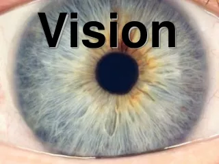
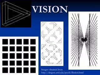
VISION. Images obtained from http://dragon.uml.edu/psych/illusion.html. Detection, Transduction, Coding. Sensory receptors : specialized receptors detect and respond to environmental stimuli Sensory Transduction : conversion of physical energy into a neural signal

VISION
E N D
Presentation Transcript
VISION Images obtained from http://dragon.uml.edu/psych/illusion.html
Detection, Transduction, Coding • Sensory receptors: specialized receptors detect and respond to environmental stimuli • Sensory Transduction: conversion of physical energy into a neural signal • Neural Coding: specific pattern of neural activity that contains information about environmental stimuli
Retinal Circuitry • Photoreceptors (rods and cones) • Horizontal cells • Bipolar cells • Amacrine cells • Retinal Ganglion Cells
Cones have better acuity due to low convergence. Rods have greater light sensitivity due to a high convergence. Convergence in the Retina
CONES (photopic system) 4 million concentrated in fovea 3 photopigments low contrast sensitivity high acuity RODS (scotopic system) 100 million outside fovea 1 photopigment high contrast sensitivity low acuity Photoreceptors
Physical Properties of Light • Wavelength is measured in nanometers and is related to the perceived characteristic of hue or color. • Intensity is related to the perceived characteristic of brightness. • Purity refers to the number of wavelengths a source of light contains and is related to the perceived characteristic of saturation.
Phototransduction • Structures of the photoreceptors • Lamella—A thin membranous disc in the outer layer of a photoreceptor. • Photopigment—A chemical molecule in the lamellae of the eye that absorbs light. • Opsin—The protein component of a photopigment. • Retinal—The lipid component of a photopigment, synthesized from Vitamin A. • Rhodopsin (rosy color before light exposure)—The photopigment in rods; consists of an opsin and a retinal.
Bleaching and Regeneration ofVisual Pigments in Rods • Insert Fig. 6.11 here
Phototransduction • Light absorbed by rhodopsin • Opsin and retinal split • Activated opsin combines with G protein to activate phosphodiesterase (PDE) • PDE breaks down cGMP to 5’-GMP • Na+ channels close, causing hyperpolarization
Retinal Coding • Bipolar cells connect photoreceptors to Retinal Ganglion Cells. • Horizontal and amacrine cells lie parallel to the retina’s surface. • Horizontal cells receive neural messages from photoreceptors and have inhibitory influences on bipolar cells. • Amacrine cells receive neural messages from the bipolar cells and inhibit both bipolar and ganglion cells.
Retinal Ganglion Cells • Retinal Ganglion Cells fire action potentials. • RGC axons form the optic nerve. • RGCs characterized by responses to light. • On ganglion cells: excited by bipolar cells in response to a light stimulus. • Off ganglion cells: excited when a light stimulus is removed and inhibited by amacrine cells in the presence of light. • On-off ganglion cell: excited by both the presence and removal of a light stimulus. • First characterized by Hartline in frogs (1938) • Kuffler characterized RGC responses in cats (1952)
Receptive fields of visual neurons: the region of the visual field where light must fall to stimulate the neuron. For any particular visual neuron, the location of its receptive field depends on the locations within the retina of the photoreceptors that provide input to that neuron. Receptive Fields
LGN Organization • PARVOCELLULAR • small cells • dorsal four layers • high spectral sensitivity • low contrast sensitivity • high spatial resolution • low temporal resolution • MAGNOCELLULAR • large cells • ventral two layers • low spectral sensitivity • high contrast sensitivity • low spatial resolution • high temporal resolution
Young-Helmholtz (trichromatic) theory Color perceptions come from a pattern of stimulation of three sets of color receptors in the eye. In 19th century, based merely on psychophysical evidence I Modern evidence: three cone types Color Coding by the Retina
Opponent-process theory—The theory that there are three receptor complexes operating in opponent fashion to yield a perception of color and brightness. This theory explains negative afterimages. Theories of Color Perception
Color Perception • Integration of Young-Helmholtz trichromatic theory and Hering’s opponent-process theory • There are different types of cones which are sensitive to different wavelengths as predicted by the trichromatic theory. • Beyond the level of photoreceptors there are different types of ganglion cells and parvocellular neurons of the LGN that seem to operate by opponent-process theory.
From Trichromatic Stimulation to Opponent-process Responding
Receptive Fields in Visual Cortex • Simple cell—A neuron in area V1 that responds to lines (edges) in a specific part of the visual field having a specific orientation. If the orientation of the line is changed, the simple cell doesn’t fire or has a drastically reduced response.
Receptive Fields in Visual Cortex • Complex cells—found in areas V1 and V2. These cells are sensitive to a line stimulus oriented in a certain direction. Unlike simple cells, the stimulus can appear in several different locations and still activate the large receptive field of the complex cell. Some cells respond to line movement in a specific direction and others respond to line movement in any direction.
Hypercomplex cells—respond to visual stimuli of a particular orientation (line-tilt) within a relatively large receptive field. However, if the line stimulus extends beyond a specific point they do not respond (end-stopped). Receptive Fields in Visual Cortex
Ocular dominance column—A column of cells in the visual cortex all having the same amount of dominance of input from either the right or left eye. Orientation column—A column of cells in the visual cortex all responding to the same orientation of a line stimulus (line-tilt). Visual Cortex OrganizationColumns of Cells
SENSORY SYSTEM ORGANIZATION • Hierarchical Organization • Multiple levels of analysis (e.g., primary, secondary, association cortex) • Functional Segregation • Functionally distinct areas specializing in different kinds of analysis (e.g., color, form, motion perception) • Parallel Processing • Simultaneous analysis of signals in different ways by multiple parallel pathways of a neural network
DORSAL STREAM projections from V1 to posterior parietal cortex processing involved in location of objects in space for guiding movement VENTRAL STREAM projections from V1 to inferior temporal lobe processing involved in object recognition Extrastriate Visual Pathways
Color Perception • Cerebral achromatopsia: inability to discriminate among different hues; caused by damage to inferior temporal cortex (V4, now called V8) of the visual association cortex. • e.g., “The Case of the Color Blind Painter” by Oliver Sacks
Form Perception • Perceptual problems with form recognition may be caused by damage within the visual association cortex even though the primary visual pathway is intact. • Visual agnosia: an inability to identify/recognize /name objects presented visually, despite normal visual acuity and object identification by other senses is otherwise normal.
Visual Agnosia • Apperceptive • Damage to ventral stream • Despite normal visual acuity, can not identify objects by sight; also can not draw objects or copy pictures by sight • Associative • Ventral and dorsal streams intact; disruption of connections between ventral stream and verbal mechanisms • Can copy objects or drawings by sight, but can not do so from memory. • Problem transferring perception to verbal mechanisms/conscious awareness.
Visual Agnosia Prosopagnosia: An impaired ability to recognize specific faces visually. Fusiform face area: The region of the inferotemporal cortex most responsible for recognition of faces.
Motion Perception • Akinetopsia: an inability to perceive movement, caused by damage to area V5 (also called MST) of the visual association cortex.
Motion Perception • Balint’s syndrome: a syndrome caused by bilateral damage to the parieto-occipital region; includes optic ataxia, ocular apraxia, and simultanagnosia. • optic ataxia: difficulty in reaching for objects under visual guidance. • ocular apraxia: difficulty in visual scanning. • simultanagnosia: difficulty in perceiving more than one object at a time.
Motion Perception • intraparietal sulcus (IPS) • The end of the dorsal stream of the visual association cortex; involved in perception of location, visual attention, and control of eye and hand movements.