Scaphoid Fractures
Scaphoid Fractures. Scaphoid Fractures. The scaphoid is the most frequently fractured carpal bone, accounting for 71% of all carpal bone fractures. Scaphoid fractures often occur in young and middle-aged adults, typically those aged 15-60 years.


Scaphoid Fractures
E N D
Presentation Transcript
Scaphoid Fractures • The scaphoid is the most frequently fractured carpal bone, accounting for 71% of all carpal bone fractures. • Scaphoid fractures often occur in young and middle-aged adults, typically those aged 15-60 years. • About 5-12% of scaphoid fractures are associated with other fractures • 70-80% occur at the waist or mid-portion • 10-20% proximal pole
Anatomy • The scaphoid lies at the radial border of the proximal carpal row, but its elongated shape and position allow bridging between the 2 carpal rows because it acts as a stabilizing rod. • The scaphoid has 5 articulating surfaces: • with the radius, lunate, capitate, trapezoid, and trapezium. • As a result, nearly the entire surface is covered by hyaline cartilage.
Blood Supply • Vessels may enter only at the sites of ligamentous attachment: • the flexor retinaculum at the tubercle, • the volar ligaments along the palmar surface, • and the dorsal radiocarpal and radial collateral ligaments along the dorsal ridge.
Blood Supply • Classically described as 3 principal arterial groups, but in more recent investigations by Gelberman and Menon described 2: • Entering dorsally • Volar side limited to tubercle
Blood Supply • The primary blood supply comes from the dorsal branch of the radial artery, which divides into 2-4 branches before entering the waist of the scaphoid along the dorsal ridge. • The branches course volar and proximal within the bone, supplying 70-85% of the scaphoid. • The volar scaphoid branch also enters the bone as several perforators in the region of the tubercle; these supply the distal 20%-30% of the bone
All studies consistently demonstrated poor supply to the proximal pole • The proximal pole is an intra-articular structure completely covered by hyaline cartilage with a single ligamentous attachment • Deep radioscapholunate ligament • Is dependent on intraosseous blood supply Blood Supply
Blood Supply • Obletz and Halbstein in their study of vascular foramina in dried scaphoids found 13% without vascular perforations and 20% with only a single small foramen proximal to the waist • Therefore postulated that atleast 30% of mid-third fracture would expect AVN of proximal pole…greater likelihood the more proximal the fracture
Pathophysiology • The primary mechanism of injury to the scaphoid bone is a fall on an outstretched hand. • A scaphoid fracture is part of a spectrum of injuries based on 4 factors: • (1) the direction of 3-dimensional loading, • (2) the magnitude and duration of the force, • (3) the position of the hand and wrist at the time of injury, and • (4) the biomechanical properties of ligaments and bones. • These factors affect the end result of the fall: distal radius fracture, ligamentous injury, scaphoid fracture, or a combination of these.
Pathophysiology • Essentially fractures of scaphoid have been explained as a failure of bone cause by compressive or tension load • Compression, as explained by Cobey and White, against concave surface by head of capitate • Position of radial and ulnar deviation thought to determine where it breaks • Fryman subjected cadaver wrists to loading and observed that: • extension of 35 degrees of less resulted in distal forearm fractures • >90degrees resulted in carpal fractures • Combination of radial deviation and wrist extension locks scaphoid within the scaphoid fossa
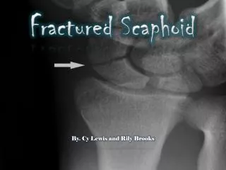
Diagnosis • Suggested by: • patient’s age, • mechanism of injury and • signs and symptoms • Imaging • Xray • CT Scan • MRI • Bone Scan
Radiography • The 4 essential views (ie, PA, lateral, supinated and pronated obliques) identify majority of fractures. • The scaphoid view is a PA radiograph with the wrist extended 30° and deviated ulnarly 20°. This view helps to stretch out the scaphoid and is also used for assessing the degree of scaphoid fracture angulation. • A clenched-fist radiograph has also been useful for visualization of the scaphoid waist.
CT Scans • CT permits accurate anatomic assessment of the fracture. • Bone contusions are not evaluated with CT, but true fractures can be excluded
MRI • T1-weighted images obtained in a single plane (coronal) are typically sufficient to determine the presence of a scaphoid fracture. • Gaebler prospectively performed MRI on 32 patients, at average of 2.8 days post injury • 100% sensitivity and specificity • In recent study Dorsay has shown that immediate MRI provides cost benefit when compared to splintage and repeat xray • False positives due MRI’s sensitivity to marrow oedema
Nuclear Imaging • Radionuclide bone scanning typically is performed 3-7 days after the initial injury if the radiographic findings are normal. • Best at 48hours, premature imaging may be obscured by traumatic synovitis • Bone scan findings are considered positive for a fracture when intense, focal tracer accumulation is identified. • Negative bone scan results virtually exclude scaphoid fracture • Teil-van studied cost effectiveness and concluded that initial xray followed by bone scan at 2 weeks if patient is still symptomatic is most effective management option • Teil-van also suggested that more sensitive and less expensive than MRI
Classification • Determining optimal treatment depends on accurate diagnosis and fracture classification • Herbert devised an alpha-numeric system that combined fracture anatomy, stability and chronicity of injury.
Herbert’s Classification • Type A (stable acute fractures) • A1: fracture of tubercle • A2: incomplete fracture • Type B (unstable acute fractures) • B1: distal oblique • B2: complete fracture through waist • B3: proximal pole fracture • B4: trans-scaphoid perilunate fracture dislocation of carpus
Herbert’s Classification • Type C (delayed union) • Type D (established non-union) • D1: fibrous union • D2: pseudarthrosis
Russe Classification • Russe classified scaphoid fractures into 3 type according to the relationship of the fracture line to the long axis of the scaphoid • Horizontal • Oblique • Vertical (unstable)
Classification according to location • A: tubercle • B: distal pole • C: waist • D:proximal pole
Management • Proximal pole • Depends on size and vascularity of fracture • Growing sentiment that most should be treated operatively because of high propensity for non-union and increased duration of immobilisation required for non-operative management • If large enough to accommodate a screw than every attempt should be made
Management • DeMaagd and Engber showed 11 of 12 patients with proximal pole fractures healed with Herbert screw • Retting and Raskin had 100% union in 17 cases with Herbert screw • If fragment too small then K-wires can be used
Management • Distal Pole • Are infrequent • Usually extra-articular with good blood supply • Best treated with short arm thumb spica for 3-6 weeks
Management of waist fractures • Most common type of fracture • High rate of delayed and non-union • With delays in treatment adversely affect results • Operative vs non-operative • Controversial
Management of waist fractures • Most stable fractures can be treated with below elbow thumb spica • Unstable fractures best treated with compression screw fixation • >1mm displacement • Fragment angulation • Abnormal carpal alignment • With advent of percutaneous techniques of cannulated screws under flouroscopic control trend towards operative management
What about the undisplaced waist fractures??? • Netherlands study: • Average time away from work 4.5 months • Saeden in prospective randomised study with 12 year follow-up compared early operative vs cast immobilisation • Return to work quicker in operative • No significant long term difference in functional outcome between 2 groups • Bond has shown return to work 7 weeks earlier and time of union 5 weeks quicker • Other papers disagree • Some surgeons published union rates of 100% with surgery(Green’s volume 1 page 721)
Complication$$ • Malunion • Malunion may lead to limited motion about the wrist, decreased grip strength, and pain. • The most frequent pattern of malunion is persistent angular deformity, or the humpback deformity. • Malunion usually can be treated with osteotomy and bone grafting to correct angular deformity and length. • Literature confusing with no comparative studies to document improvement in hand function
Complication$$ • Delayed union and non-union • Delayed union is incomplete union after 4 months of cast immobilization. • Non-union is an unhealed fracture with smooth fibrocartilage covering the fracture site. • About 10-15% of all scaphoid fractures do not unite. • Some degree of delayed union or non-union occurs in nearly all proximal pole fractures and in 30% of scaphoid waist fractures