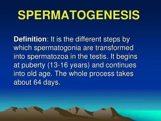
SPERMATOGENESIS
SPERMATOGENESIS. Mitosis. 24 primary spermatocytes. Spermatogenic cycle. Mitosis. Mitosis. Mitosis. Meiosis II. Meiosis I. 2A 1. A 1. 96 spermatids. 6B 1. 3A 2. 12B 2. 48 secondary spermatocytes. Spermiogenesis = Spermateleosis = Spermatozoan metamorphosis. Mitosis.

SPERMATOGENESIS
E N D
Presentation Transcript
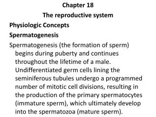
Mitosis 24 primary spermatocytes Spermatogenic cycle Mitosis Mitosis Mitosis Meiosis II Meiosis I 2A1 A1 96 spermatids 6B1 3A2 12B2 48 secondary spermatocytes Spermiogenesis = Spermateleosis = Spermatozoan metamorphosis Mitosis 96 mature spermatozoa The following is an example of how the number of spermatozoa is increased by repetitive mitotic divisions of spermatogonial cells followed by the two meiotic divisions. There are actually more than 4 types of spermatogonia, so the actual number of mature spermatozoa originating from the initial division of a type A1 spermatogonium is actually greater than 96. A1 First meiotic division lasts several weeks in humans Reductional division Second meiotic division takes about 8 hours in humans Equational division An entire spermatogenic cycle in humans takes about 64 days. The maturing spermatids remain attached by cytoplasmic bridges as they mature => syncytium
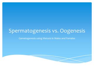
Let’s think about meiosis: 1. Where did the chromosomes come from? 2. Why are there pairs of chromosomes? 3. How must the chromosomes segregate if you’re going to have sexual reproduction? What do you have to end up with in he gametes? 4. How would you accomplish this in two divisions?
I Homologous Gene Loci X X M I X X I P P M P Replication Replication Replicated Homologous Gene Loci M X Replicated Chromosome Dyad - Two Chromatids Homologous Pair Replicated Homologous Pair CHROMOSOME TERMINOLOGY Single Chromosome Monad Synapsis Tetrad = Two Synapsed replicated Homologues
M P M P M M P P Genetic content vs Chromosome number Diploid genetic content - 2n - the 2 represents the fact that each gene locus can contain a maximum of 2 different gene alleles (e.g., one dominant and one recessive). “n” is the total number of homologous gene loci in the genome (a very big number). Diploid chromosome number - 2c - the 2 represents the fact that there are 2 chromosomes in each homologous pair. “c” is the total number of homologous pairs of chromosomes. Meiosis Genetic Content (n) and Chromosome Number (c)
Leptotene Chromosomes start to condense, dyads (replicated chromosomes) of homologous pairs first become visible as linear strings of DNA Zygotene Chromosomes condense further. Dyads of homologous pairs of chromosomes pair-up and their chromatids start to undergo synapsis. Pachytene Synapsis is completed with synapsed chromosomes forming tetrads. Cross-over takes place. Synapsed chromosomes thicken up (pachy - thick). Diplotene Chromosomes condense further, Desynapsis begins. Chromatids of homologous pairs of chromosomes remain connected at chiasmata where cross-over may have occurred. Diakinesis Chromosomes condense even further. Separating tetrads form strange shapes like crosses, fish, infinity signs as chiasmata move toward the ends of the paired chromatids of the homologous chromosomes (called terminalization). Eventually desynapsis is completed and the homologues separate from one and other completely and metaphase begins. First meiotic prophase:
Spermatogenesis in Vertebrates Figure on Page 89 of your text
Spermatogenesis in Vertebrates Figure of rat testis in digital lab manual
Spermatogenesis in Vertebrates Figure of human testis in digital lab manual
Spermiogenesis 1. Nucleus condenses (chromosomes condense and nuclear sap is removed) 2. Flagellum develops 3. Spermatocyte elongates 4. Acrosome formed from golgi body 5. Mitochondria aggregate around base of forming flagellum 6. Mitochondria fuse to form supermitochondrion (in humans) 7. Most of cytoplasm is shed and phagocytosed by sertoli cell (tubulobulbar processes)
1. Nucleus condenses (chromosomes condense and nuclear sap is removed) 2. Flagellum develops 3. Spermatocyte elongates 4. Acrosome formed from golgi body 5. Mitochondria aggregate around base of forming flagellum 6. Mitochondria fuse to form supermitochondrion (in humans) 7. Most of cytoplasm is shed and phagocytosed by sertoli cell (tubulobulbar processes)
Sertoli cell function 1. Remove excess cytoplasm from developing spermatid - tubulobulbar processes 2. Move spermatids toward the lumen of the seminiferous tubules - ectoplasmic specializations 3. Nurture and mediate maturation of spermatids 4. Segregate groups of developing gametes 5. Secrete fluid to transport sperm in reproductive tract 6. Secrete hormones and other factors a. Embryonic - anti-mullarian hormone b. Adult (1) inhibin (inhibits FSH production) (2) estrogen - may act to inhibit GnRH production by basal hypothalamus (3) Otherfactor (not a hormone) - androgen binding protein (helps transport androgens from interstitial fluid into seminiferous tubule - promotes spermatogenesis) http://education.vetmed.vt.edu/Curriculum/VM8054/Labs/Lab27/EXAMPLES/EXSERTOL.HTM