Download

1 / 10
110 likes | 348 Vues
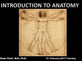
Anatomy Lab Slides. Anatomy of the hand. Kaan Yücel M.D., Ph.D. 15.February.2012 Wednesday. 1. What are flexor retinaculum & carpal tunnel?. The carpal tunnel formed anteriorly at wrist by a deep arch formed by carpal bones & f lexor retinaculum (transverse carpal ligament).

E N D
AnatomyLab • Slides • Anatomy • of the hand • Kaan Yücel M.D., Ph.D • 15.February.2012 Wednesday
1. What are flexor retinaculum & carpal tunnel? The carpal tunnelformed anteriorly at wrist by a deep arch formed by carpal bones & flexor retinaculum (transverse carpal ligament) Flexor digitorum/superficialis Flexor pollicis longus Median nerve Pass through the carpal tunnel
2. What is extensor retinaculum (dorsal carpal ligament)? The extensor tendons pass into the hand in six compartments defined by an extensor retinaculum: • extensor digitorum & extensor indicis posterior surface of the wrist extensor carpi ulnaris & extensor digiti minimi medial side of the wrist • abductor pollicis longus & extensor pollicis brevis • extensor carpi radialis longus & extensor carpi radialis brevis • extensor pollicis longus • through three compartments on the lateral surface of the wrist.
3. What is palmar aponeurosis? A triangular condensation of deep fascia that covers the palm and is anchored to the skin in distal regions. Continuous with the palmaris longus tendon, when present; otherwise, anchored to the flexor retinaculum.
4. What are extensor hoods for? Tendons of the extensor digitorumextensor pollicis longus musclesexpand over the proximal phalanges to form "extensor hoods" or "dorsal digital expansions". Tendons of the extensor digiti minimi, extensor indicis, extensor pollicis brevisjoin these hoods.
5. Which are the intrinsic muscles of the hand? Palmaris brevis Thenar muscles Abductor pollicis brevis Abductor digiti minimi Opponens pollicis Hypothenar muscles Flexor digiti minimi Adductor pollicis Flexor pollicis brevis Lumbrical Interossei
6. ....arteries of the hand? Superficial palmar arch Deep palmar arch
7. ....veins of the hand? Cephalic veinoriginates from lateral side of dorsal venous network. Basilic vein originates from medial side of dorsal venous network.
8. ...carpal tunnel? Homework: • 1. Which structures pass through the carpal tunnel and their anatomical relationships with each other in the tunnel? • 2. The incidence of carpal tunnel syndrome in the world and/or in Turkey? • 3. The risk factors, higher in whom? Any gender disperancies in its incidence? Please send answers to yeditepeanatomy@yahoo.com