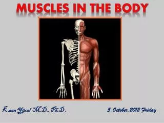
Kaan Yücel M.D., Ph.D
600 likes | 916 Vues
ARM. Kaan Yücel M.D., Ph.D. 4.January.2012 Wednesday. Arm is the region of the upper limb between the shoulder and the elbow. The superior aspect of the arm communicates medially with the axilla.

Kaan Yücel M.D., Ph.D
E N D
Presentation Transcript
ARM • Kaan Yücel M.D., Ph.D • 4.January.2012 Wednesday
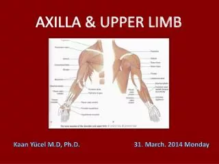
Arm is the region of the upper limb between the shoulder and the elbow. • The superior aspect of the arm communicates medially with the axilla. • Inferiorly, a number of important structures pass between arm & forearm through cubital fossa, positioned anterior to the elbow joint.
The arm is divided into two compartments by medial& lateral intermuscular septa pass from each side of the humerus to the outer sleeve of deep fascia that surrounds the limb.
The anterior compartment of the arm contains muscles that predominantly flex the elbow joint; the posterior compartment contains muscles that extend the joint. Major nerves and vessels supply and pass through each compartment. The chief action of both groups is at the elbow joint, but some muscles also act at the glenohumeral joint.
Two types of movement occur between the arm and the forearm at the elbow joint: flexion-extension and pronation-supination. The muscles performing these movements are clearly divided into anterior and posterior groups, separated by the humerus and medial and lateral intermuscular septae.
Anterior compartment of the arm coracobrachialis, brachialis, and biceps brachii muscles innervated predominantly by musculocutaneous nerve. Posterior compartment triceps brachii muscle innervated by radial nerve.
Coracobrachialis Anelongated muscle in the superomedial part of the arm. Useful landmark for locating other structures in the arm musculocutaneous nerve pierces it distal part of its attachment indicates location of nutrient foramen of the humerus
Coracobrachialis Extends from tip of coracoid process of the scapula to medial side of the midshaft of humerus.
Coracobrachialis 1. helps flex and adduct the arm 2. stabilize the glenohumeral joint. With deltoid + long head of triceps a shunt muscle, resisting downward dislocation of the head of the humerus, as when carrying a heavy suitcase. Median nerve and/or brachial artery may run deep to coracobrachialis and be compressed by it.
Coracobrachialis It passes through the axilla and is penetrated and innervated by the musculocutaneous nerve.
Proximal attachment of this fusiform muscle usually has two heads • (bi, two + L. caput, head). • short head originates from coracoid process in conjunction with the coracobrachialis • long head originates as a tendon from supraglenoid tubercle of scapula. Biceps brachii
Biceps brachii The tendon of the long head passes through the shoulder joint superior to the head of the humerus, and enters the arm. In the arm, together with the muscle belly of the short head, overlies the brachialis muscle. Two heads a single tendon, inserts onto radial tuberosity.
Biceps brachii Transverse humeral ligamentconverts the intertubercular grooveinto a canal& holds the tendon of long head of biceps in the groove.
Biceps brachii Triangular membranous band, bicipital aponeurosis, runs from the biceps tendon across the cubital fossa and merges with antebrachial (deep) fascia covering the flexor muscles in the medial side of the forearm. Affords protection for these & other structures in the cubital fossa. Helps lessen the pressure of the biceps tendon on the radial tuberosity during pronation & supination of the forearm.
Biceps brachii • “Three-joint muscle,” crossing & capable of effecting movement at the • Glenohumeral joint • Elbow joint • Radio-ulnar joint primarily acts at the latter two. • Powerful flexor of the forearmat the elbow joint • Most powerful supinator of the forearm when elbow joint is flexed. • Because two heads of biceps brachii muscle cross the glenohumeral joint, the muscle can also flex the glenohumeral joint.
Biceps brachii Its action and effectiveness are markedly affected by the position of the elbow and forearm. Elbow extended a simple flexor of the forearm
Biceps brachii Elbow flexion approaches 90° and more power is needed against resistance, capable of 2 powerful movements, depending on the position of the forearm. 1) Elbow is flexed close to 90° & forearm supinated: biceps most efficient in producing flexion. 2) Forearm pronated, biceps primary (most powerful) supinator of forearm.
Biceps brachii Innervated by the musculocutaneous nerve. A tap on the tendon of biceps brachii at the elbow is used to test predominantly spinal cord segment C6.
Brachialis Originates from the distal half of the anterior aspect of the humerus and from adjacent parts of the intermuscular septa, particularly on the medial side. It lies beneath the biceps brachii muscle and converges to form a tendon, which attaches to the tuberosity of the ulna & coronoid process of ulna. Its distal attachment covers the anterior part of the elbow joint.
Brachialis Main flexor of the forearm The only pure flexor, producing the greatest amount of flexion force Always contracts when the elbow is flexed and is primarily responsible for sustaining the flexed position. Because of its important and almost constant role, it is regarded as the workhorse of the elbow flexors.
Brachialis Innervation predominantly by musculocutaneous nerve. A small component of the lateral part is innervated by the radial nerve.
Triceps brachii Large fusiform muscle in the posterior compartment of the arm. The only muscle of the posterior compartment • 3 heads originate from: • long head infraglenoid tubercle of scapula • medial head & lateral head posterior surface of humerus, superior to radial groove3 heads converge to form a large tendon, inserts on • superior surface of the olecranon of the ulna
Triceps brachii Because its long head crosses the glenohumeral joint, the triceps helps stabilize the adducted glenohumeral joint by serving as a shunt muscle, resisting inferior displacement of the head of the humerus. The long head also aids in extension and adduction of the arm, but it is actually the least active head. Medial head : workhorse of forearm extension, Lateral head : strongest but is recruited into activity primarily against resistance.
Triceps brachii Innervation of by branches of the radial nerve. A tap on the tendon of triceps brachii tests predominantly spinal cord segment C7.
Brachial artery The major artery of the arm Found in the anterior compartment Continuation of axillary artery at the lower border of teres major Terminates distal to the elbow joint, opposite to neck of radius dividing into radial & ulnar arteries.
Brachial artery Relatively superficial and palpable throughout its course. Lies anterior to triceps & brachialis. As it passes inferolaterally, accompanies the median nerve.
Brachial artery Proximal arm lies on the medial side. Distal arm, it moves laterally. Named Branches Superior ulnar collateral artery Inferior ulnar collateral artery contribute to a network of arteries around the elbow joint. Profunda brachii artery Nutrient arteries to the humerus
Brachial artery Deep artery of the arm (L. arteria profunda brachii) Largest branch & most superior origin Accompanies radial nerve along the radial groove Terminates by dividing into middle & radial collateral arteries, participate in the periarticular arterial anastomoses around the elbow.
Brachial artery Deep artery of the arm (L. arteria profunda brachii) Largest branch & most superior origin Accompanies radial nerve along the radial groove Terminates by dividing into middle & radial collateral arteries, participate in the periarticular arterial anastomoses around the elbow.
Two sets of veins of the arm superficial and deep, anastomose freely with each other. Superficial veins in the subcutaneous tissue, Deep veins accompany the arteries. 2 main superficial veins of the arm cephalicand basilic veins. Cephalic vein ascends in the superficial fascia on the lateral side of the biceps and drains into the axillary vein.
Paired deep veins, collectively constite brachial vein, accompany the brachial artery. The pulsations of the brachial artery help move the blood through this venous network. The brachial vein begins at the elbow by union of the accompanying veins of the ulnar and radial arteries and ends by merging with the basilic vein to form the axillary vein.
NERVES IN THE ARM
4 main nerves pass through the arm: • Median • Ulnar • Musculocutaneous • Radial
Musculocutaneous nerve Leaves the axilla and enters the arm by passing through the coracobrachialis muscle. Passes diagonally down the arm between biceps brachii & brachialis.
Musculocutaneous nerve • After giving rise to motor branches in the arm, it emerges laterally to the tendon of the biceps brachii muscle at the elbow, • Becomes truly subcutaneous to course initially w/cephalic vein • Provides: • motor innervation to all muscles @ anterior compartment of the arm; • sensory innervation to skin @ lateral surface of the forearm.
Median nerve Enters the arm from axilla @ inferior margin of teres major muscle. Passes vertically down the medial side of arm in the anterior compartment Related to brachial artery throughout its course: No major branches in the arm, or in the axilla.