ARTICULATIONS IN THE BODY
600 likes | 943 Vues
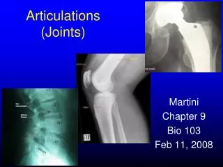
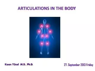
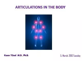
ARTICULATIONS IN THE BODY. Kaan Yücel M.D., Ph.D . 5. March . 201 3 Tuesday. Arthrology Greek a rqron joint – logy. science concerned with the anatomy , function, dysfunction and treatment of joints. Joints (articulations)

ARTICULATIONS IN THE BODY
E N D
Presentation Transcript
ARTICULATIONS IN THE BODY Kaan Yücel M.D., Ph.D. 5. March. 2013 Tuesday
Arthrology Greek a rqron joint –logy • science concerned with the • anatomy, function, dysfunction and treatment of joints.
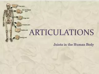
Joints (articulations) • unions or junctions between two or more bones or rigid parts of the skeleton • Whetheror not movement occurs still called a joint. • Some joints have no movement • Others only slight movement • Some freely movable
according to the tissues that lie between the bones: Fibrous joints Cartilaginous joints Synovial joints Classification of Joints
Fibrous joints • Bones are united by fibrous tissue. • Suturesof the cranium
Fibrous joints • Syndesmosistype of fibrous joint • unites the bones with a sheet of fibrous tissue • either a ligament or a fibrous membrane • partially movable • The interosseous membrane in the forearm is a sheet of fibrous tissue that joins the radius and ulna in a syndesmosis.
Fibrous joints • Syndesmosistype of fibrous joint • unites the bones with a sheet of fibrous tissue • either a ligament or a fibrous membrane • partially movable • The interosseous membrane in the forearm is a sheet of fibrous tissue that joins the radius and ulna in a syndesmosis.
Cartilaginous joints Bones are united by hyaline cartilage or fibrocartilage.
Cartilaginous joints Pimarycartilaginous joints-synchondroses hyaline cartilage- growth of a bone duringearly life Secondary cartilaginous joints-symphyses strong, slightly movable joints united by fibrocartilage
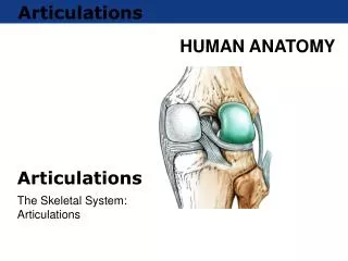
Synovial joints Most common type of joints Bones united by a joint capsule enclosing an articular cavity. Providefree movement between the bones they join. • Joint cavity • potential space • contains lubricating synovial fluid, secreted by the synovial membrane. • Articular cartilage • articular surfaces are covered by hyaline cartilage • Articular capsule • surrounds the joint and formed of two layers.
Articular capsule: • surrounds the joint • two layers. • Fibrous capsule • Synovial membrane Some synovial joints have other distinguishing features, such as a fibrocartilaginousarticular disc or meniscus, which are present when the articulating surfaces of the bones are incongruous.
Ligaments • a cordorband of connectivetissueunitingtwostructures. • Articularcapsulesareusuallystrengthenedbyarticularligaments. • Connect thearticulatingbonestoeachother. • limit theundesiredand/orexcessivemovements of thejoints.
Articulardisc: Help toholdthebonestogether. Labrum: A fibrocartilaginous ring whichdeepensthearticularsurfaceforone of thebones.
Bursa • Flattenedsacsthatcontainsynovialfluidtoreducefriction. • Wallsare separated by a film of viscous fluid. • Foundwherever tendons rub against bones, ligaments, or other tendons.
Stability of Joints Negativepressure within the joint cavity Shape, size, and arrangement of the articular surfaces Ligaments Tone of the muscles around the joint
Jointvasculatureandinnvervation • Joints receive blood from articular arteries that arise from the vessels around the joint. • Articular veins are communicating veins that accompany arteries (L. venae comitantes) and, like the arteries, are located in the joint capsule, mostly in the synovial membrane. • Joints have a rich nerve supply provided by articular nerves with sensory nerve endings in the joint capsule.
Types of synovial joints according to shape of articulating surfaces- type of movement they permit Plane joints uniaxialjoints- glidingorsliding acromioclavicular joint 2. Hinge joints uniaxialjoints- flexion & extension knee & elbowjoints
Types of synovial joints 3. Saddle joints biaxialjoints- flexion & extension, abduction & adduction carpometacarpal joint at the base of the 1st digit (thumb) 4. Condyloid (ellipsoid type) biaxialjoints- flexion & extension, abduction & adduction metacarpophalangeal joints (knuckle joints) radiocarpal joint (wrist)
Types of synovial joints 5. Ball and socket joints (spheroidaljoints) multiple axes and planes: flexion and extension, abduction and adduction, medial and lateral rotation, and circumduction hip & shoulderjoints
Types of synovial joints 6. Pivot joints uniaxialjoints- rotationaround a centralaxis proximal& distal radioulnar joints
TEMPOROMANDIBULAR JOINT a modified hinge type of synovial joint Movements • gliding (translation) • small degree of rotation (pivoting) • flexion (elevation) • extension (depression)
TEMPOROMANDIBULAR JOINT • mandibular fossa &articular tubercle of temporal bone • head of the mandible • articular disc of the TMJ
JOINTS OF THE VERTEBRAL COLUMN • The vertebral column in an adult typically consists of 33 vertebrae arranged in five regions: 7 cervical, 12 thoracic, 5 lumbar, 5 sacral, and 4 coccygeal. • Joints of the vertebral bodiessymphyses (secondary cartilaginous joints) • Joints of the vertebral arches(facetjoints) • Craniovertebral(atlanto-axial and atlanto-occipital) joints • Costovertebraljoints • Sacroiliac joints
Jointsof the vertebral bodies • designed for weight-bearing and strength • Twovertebraeconnected by intervertebral (IV) discs and ligaments. • Discsprovide strong attachments between the vertebral bodies.
1. anulusfibrosus(L. anus, a ring) bulging fibrous ring forming the circumference of the IV disc 2. anterior longitudinal ligament covers and connects the anterolateral aspects of the vertebral bodies and IV discs. 3. posterior longitudinal ligament runs within the vertebral canal along the posterior aspect of the vertebral bodies.
Jointsof the vertebral arches • between superior &inferior articular processes of adjacent vertebrae • The adjacent vertebral arches are joined by broad, pale yellow bands of elastic tissue called the ligamentaflava(L. flavus, yellow). PLANE TYPE SYNOVIAL JOINT GLIDING MOVEMENTS
MOVEMENTS OF THE VERTEBRAL COLUMN The range of movement varies according to the region and the individual. The mobility results primarily from the compressibility and elasticity of the intervertebral discs. Movements by the vertebral column include flexion, extension, lateral flexion, rotation, and circumduction.
Craniovertebraljoints A. atlanto-occipitaljoints between atlas (C1 vertebra), & occipital bone of thecranium B. atlanto-axialjoints between atlas &axis(C2 vertebra) Theirdesigngives a widerrange of movementthan in the rest of thevertebralcolumn.
Craniovertebraljoints Atlanto-occipital joints nodding of the head, such as the flexion and extension of the head approval “yes” movement 3 Atlanto-axial articulations 2(right and left) lateral atlantoaxialjoints 1median atlantoaxial joint. head turned from side to side, “no” movement
JOINTS OF THE UPPER LIMB Sternoclavicularjoint (SC) sternalend of the clavicle articulates with manubrium & 1st costal cartilage Theonlyarticulationbetweenupperlimb& axialskeleton. During full elevation of the limb, clavicle is raised to 60° angle.
Glenohumeral(shoulder) joint permits a wide range of movement; mobility makes the joint relatively unstable. Humeralhead articulates w/ glenoidcavity of the scapula deepened slightly but effectively by the ring-like, fibrocartilaginousglenoid labrum (L., lip).
Glenohumeral(shoulder) joint more freedom of movement than any other joint in the body results from the laxity of its joint capsule & large size of the humeral head compared with the small size of the glenoid cavity. movements around three axes flexion-extension, abduction-adduction, rotation (medial and lateral) of the humerus, circumduction
ElbowJoint • located inferior to the epicondyles of the humerus • humeroulnar&humeroradialarticulations
collateral ligaments of the elbow joint • strong triangular bands • medial and lateral thickenings of the fibrous layer of the joint capsule • Radialcollateral ligament • Ulnarcollateral ligament • Flexion and extension occur at the elbow joint. • Intratendinous olecranon bursa • Subtendinous olecranon bursa • Subcutaneous olecranon bursa
Proximal(superior) radio-ulnar joint allows movement of the head of the radius on the ulna Radialhead is held in position by the anular ligament of the radius. Distal(inferior) radio-ulnar joint The radius moves around the relatively fixed distal end of the ulna.
Wrist(radiocarpal) joint ulna does not participate in the wrist joint. Distalend of the radius & articular disc of the distal radio-ulnar joint articulate with proximal row of carpal bones, except for the pisiform. Flexion Extension Abduction Adduction radialdeviation-ulnardeviation Circumduction
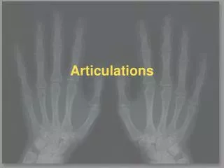
Intercarpal joints interconnect the carpal bones. Carpometacarpaljoints Intermetacarpaljoints Metacarpophalangeal joints Interphalangealjoints
JOINTS OF THE LOWER LIMB articulations of the pelvic girdle lumbosacral joints, sacroiliac joints, and pubic symphysis hip joints knee joints tibiofibular joints ankle joints foot joints
JOINTS OF THE PELVIS Pubicsymphysis interpubicdisc& surroundingligaments unitethebodies of thepubicbones in themedianplane. Lumbosacraljoints L5 and S1 vertebrae articulate Sacrococcygeal joint
Feature 1: Connection between lower limb & pelvic girdle Feature 2: 2nd most movable after the shoulder joint Synovial Joint Type: Ball and socket (Head of the femur & acetabulum) Weight transfer: To the heads and necks of the femurs acetabular labrum (L. labrum, lip) fibrocartilaginousrim attached to the margin of acetabulum, increasing acetabulararticular area by nearly 10%.
Ligaments Transverse acetabular ligamentcontinuation of acetabular labrum 3 intrinsic ligaments Iliofemoral ligamentanteriorly and superiorly , strongest ligament of the body Pubofemoral ligament anteriorly and inferiorly Ischiofemoral ligament posteriorly Ligament of the head of the femur
MOVEMENTS OF HIP JOINT • Flexion-extension • Abduction-adduction • Medial-lateral rotation • Circumduction
KNEE JOINT • Feature 1: Largest & most superficial joint • Feature 2: Hing movements (Ext/Flex) combined with gliding & rotation • Synovial Joint Type: Hinge • The knee joint consists of three articulations: • 2 femorotibial articulations (lateral and medial) • between lateral & medial femoral and tibial condyles • One intermediate femoropatellar articulation • between patella & femur • No fibula involvment in the knee joint.
KNEE JOINT The stability of the knee joint depends on (1) strength & actions of the surrounding muscles and their tendons (2) ligaments that connect the femur and tibia. muscles are most important. the most important muscle in stabilizing the knee joint quadriceps femoris.
Extracapsularligaments Patellar ligament Fibular (Lateral) collateral ligament Tibial (Medial) collateral ligament Oblique popliteal ligament Arcuate popliteal ligament
INTRA-ARTICULAR LIGAMENTS Cruciate ligaments & menisci Anterior cruciate ligament(ACL) Posterior cruciate ligament(PCL)