Download
1 / 86
860 likes | 1.28k Vues
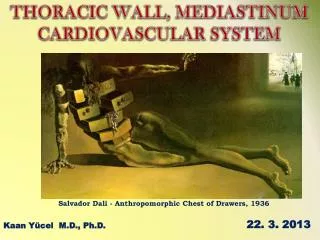
THORACIC WALL, MEDIASTINUM CARDIOVASCULAR SYSTEM. Salvador Dali - Anthropomorphic Chest of Drawers, 1936 . 22. 3. 2013. Kaan Yücel M.D., Ph.D. 1. THORAX. the part between the neck and the abdomen. Chest X-ray. 1.1. REGIONS/T E RMS. Thoracic cavity cavity between neck and abdomen

E N D
THORACIC WALL, MEDIASTINUM CARDIOVASCULAR SYSTEM Salvador Dali - Anthropomorphic Chest of Drawers, 1936 22. 3. 2013 Kaan Yücel M.D., Ph.D.
1. THORAX the part between the neck and the abdomen Chest X-ray
1.1. REGIONS/T ERMS Thoracic cavity cavity between neck and abdomen protected by the thoracic wall. Thoracic wall bounds the thoracic cavity. formed by the skin, bones, fasciae, and muscles. Thoracic cage bony portion of the thoracic wall thoracic skeleton
1.2. SURFACES OF THE THORAX STERNUM & COSTAL CARTILAGES anteriorly 12 THORACIC VERTEBRAE & POST. RIBS posteriorly RIBS & INTERCOSTAL SPACES laterally Posterior surface 12 thoracic vertebræ& posterior parts of the ribs Anterior surface sternum &costal cartilages Lateral surfaces ribs, separated by the intercostal spaces
1.3. BOUNDARIES OF THE THORAX • Superior • Jugular notch • Sternoclavicular joint • Superiorborder of clavicle • Acromion • Spinousprocesses of C7 • Inferior • Xiphoid process • Costal arch • 12th and 11th ribs • VertebraT12
1.4. CONTENTS OF THE THORAX Organs of the cardiovascular, respiratory, digestive, reproductive, immune, and nervous systems
2. THORACIC WALL • thoracic cage (skeleton) • muscles between the ribs • skin • subcutaneous tissue • muscles, and fascia covering its anterolateral aspect. • The mammary glands of the breasts lie within the subcutaneous tissue of the thoracic wall.
2.1. Functions of the thoracic wall • Protectsvital thoracic and abdominal organs • Resiststhe negative (sub-atmospheric) internal pressures generated by the elastic recoil of the lungs and inspiratory movements. • Providesattachmentfor and support the weight of the upper limbs. • Provides theoriginsof many of the muscles that move and maintain the position of the upper limbs relative to the trunk. • Provides attachments for muscles of the abdomen, neck, back, and respiration.
3. Skeleton of THEThoracic Wall • 12 pairs of ribs and associated costal cartilages • 12 thoracic vertebrae and the intervertebral (IV) discs interposed between them • Sternum
4. THORACIC APERTURES ‘Thoracic inlet’ ‘Thoracicoutlet’
4.1. Superior thoracic aperture “doorway” between the thoracic cavity and the neck and upper limb • bounded: • Posteriorly vertebra T1 • Laterally 1st pair of ribs and their costal cartilages • Anteriorlysuperior border of the manubrium • Trachea • Esophagus • nerves, and vessels that supply and drain the head, neck, and upper limbs.
4.2. Inferior thoracic aperture By closing the inferior thoracic aperture, the diaphragm separates the thoracic and abdominal cavities almost completely. • bounded: • Posteriorly12th thoracic vertebra • Posterolaterally11th and 12th pairs of ribs • Anterolaterallyjoined costal cartilages of ribs 7-10costal margins • Anteriorlyxiphisternal joint
5. JOINTS OF THE THORACIC WALL 1. Costotransverse joints between tubercle of a rib & transverse process of its own vertebra 2. Sternocostal joint between the sternum and costal cartilages 3. Costachondralisjoint between the rib and costal cartilage 4. Intercondraljoints Synovial joints between the costal cartilages of 6th and 7th, 7th and 8th, & 8th and 9th ribs. 5. Sternal Joints between the manubrium, body, xiphoid process of the sternum.
6. Muscles of the Thoracic Wall • Serratusposterior Levatorcostarum • Intercostal muscles(External, internal and innermost) • Subcostal Transverse thoracic
6. Muscles of the Thoracic Wall intercostal muscles three flat muscles found in each intercostal space external intercostal muscles most superficial internal intercostal muscles between external & innermost muscles
6. Muscles of the Thoracic Wall external intercostal muscles extend from the inferior margins of the ribs above to the superior margins of the ribs below pass obliquely anteroinferiorly
6. Muscles of the Thoracic Wall internalintercostal muscles most active during expiration between most inferior lateral edge of costal grooves of the ribs above, to the superior margins of ribs below in the opposite direction to those of the external intercostal muscles obliquely posteroinferiorly Attachmenttointerosseuspartsmovedown duringexpiration!
6. Muscles of the Thoracic Wall innermostintercostal muscles least distinct of the intercostal muscles same orientation as the internal intercostals neurovascular bundles (V.A.N.) in the costal grooves in a plane between innermost& internal intercostal muscles.
6. Muscles of the Thoracic Wall transversusthoracis muscles on the deep surface of the anterior thoracic wall & @ same plane as innermost intercostals lie deep to the internal thoracic vessels secure these vessels to the wall.
6. Muscles of the Thoracic Wall subcostales @ same plane as innermost intercostals Fibersparallel the course of the internal intercostal muscles. Extendfrom the angle of the ribs to more medial positions on the ribs below.
6.1. Accessory muscles of respiration • upper accessory muscles assist with inspiration. • upper chest, and abdominal muscles assist with expiration.
6.1. Accessory muscles of respiration Scalenemuscles elevate, fix&expanding the upper chest during inspiration. Sternocleidomasteoidmuscles raise the sternum expand the chest’s A-P& longitudinal dimensions.
6.1. Accessory muscles of respiration Axioappendicularmuscles act primarily on the upper limbs. pectoralismajor Attachmenttofirst seven costalcartilages pectoralisminor Attachmentto anteriorsurfaces of the 3rd-5th ribs inferior part of the serratusanteriorAttachmentto lateral surfaces of upper 8-9 ribs & deep fascia overlying related intercostal spaces help elevate the ribs to expand the thoracic cavity.
6.1. Accessory muscles of respiration Trapeziusraises the thoracic cage. Attachmenttospinousprocesses of thethoracalvertebrae
6.1. Accessory muscles of respiration Expiration Abdominalmuscles pull the lower chest down depress the lower limbs compress the abdominal content & exerts pressure on chest.
7. MOVEMENTS OF THE THORACIC WALL One of the principal functions of the thoracic wall and the diaphragm is to alter the volume of the thorax and thereby move air in and out of the lungs. During breathing, the dimensions of the thorax change in vertical, lateral, and A-P directions. Diaphragm contracts Depression Diaphragmrelaxes Elevation (duringpassiveexpiration) Elevation&depression of the ribs
Diaphragmconracts; thoraciccavitydescends, compressingtheabdominalviscera expiration(backtoneutralposition) Lungsproducessub-atmosphericpressure As a result, the diaphragm ascends, diminishing the vertical dimension. Verticaldimension inspiration
A-P dimension intercostal muscles contract pump-handle movement Movement of the ribs (primarily 2nd-6th) at the costovertebral joints causes the anterior ends of the ribs to rise + Slightelevation in A-P of thesternum .
Transversedimension intercostal muscles contract, raising the middle (lateral-most parts) of the ribs (especially the lower ones) bucket-handle movement
9. VASCULATURE OF THE THORACIC WALL Mainlyposterior and anterior intercostal arteries pass between adjacent ribs in intercostal spaces.
9.1. Arteries of THE Thoracic Wall • Arterialsupply to the thoracic wall derives from • Thoracic aorta • [posterior intercostal & subcostal arteries] • Subclavianartery • [internal thoracic & supreme intercostal arteries] • Axillary artery • [superior &lateral thoracic arteries]
intercostal arteries • course through the thoracic wall between the ribs. • Eachintercostalspace is suppliedby • a largeposteriorintercostalartery • smallpair of anteriorintercostalarteries • Exceptionlasttwointercostalspaces
Anterior intercostal arteries (paired) directlyorindirectlyfrominternalthoracicartery 1st-6th (from internal thoracic artery) 7th-9th (from musculophrenic-branch of internal thoracic) Posterior intercostal arteries (large, unpaired) 1st-2nd (from supreme intercostal artery- branch of costocervical trunk) 3rd-11th (from thoracic aorta) Subcostal artery (from thoracic aorta)
internal thoracic artery • Veryfirstbranch of thesubclavianartery • Passes vertically through the superior thoracic aperture • Liesposterior to the costal cartilages of the upper 6 ribs • Lies1 cm lateral to the sternum. @ level of 6th intercostal space, divides into 2 terminal branchessuperiorepigastricartery& musculophrenicartery
9.2. Veins of THE Thoracic Wall Veins Artery V.A.N. Nerve Most posterior intercostal veins (4-11) end @ azygos/hemiazygos venous system conveys venous blood to SVC. 1st posteriorintercostalveinsright & leftbrachiocephalicveins 2nd & 3rd (sometimes 4th) form superior intercostal vein. Rightsuperior intercostal vein final tributary of azygos vein Leftsuperior intercostal vein empties into left brachiocephalic vein.
10. NERVES OF THE THORACIC WALL 12 pairs of intercostal nerves anterior rami of spinal nerves T1 to T11 lie in the intercostal spaces between adjacent ribs. Anteriorramus of spinal nerve T12 (subcostal nerve) inferior to rib XII.
Near the angles of the ribs, the nerves pass between internal intercostal & innermost intercostal muscles. • V.A.N. • Neurovascular bundles sheltered by the inferior margins of the overlying rib.
10.3. Dermatomes Skin areasupplied by a segment of the spinal cord Through its posterior ramusand the lateral and anterior cutaneous branches of its anterior ramus, most thoracic spinal nerves (T2-T12) supply a strip-like dermatome of the trunkextending from the posterior median line to the anterior median line. T2- Sternal angle T4- Nipple T6- Xiphoid process T8- Costal arch T10-Umbliculus T12-Midpoint between umbilicus and symphysis pubis
11. breasts Reproduction, backpain Aesthetics, and breast cancer Mammaryglands &associated skin -connective tissues. modified sweat glands in the superficial fascia anterior to the pectoral muscles and the anterior thoracic wall.
11. breasts Mammaryglands: Seriesof ducts and associated secretory lobules. Form15 to 20 lactiferous ductsopen nipple. Nippleis surrounded by a circular pigmented area of skin areola(L. small area).
11.1. Female Breasts NON-LACTING WOMEN – PREDOMINANT COMPONENT: FAT LACTING WOMEN- PREDOMINANT COMPONENT: GLANDULAR TISSUE The breast rests on a bed extends transversely from lateral border of the sternum mid-axillary line vertically from the 2nd through 6th ribs
Lymphaticdrainage of thebreast 75% (lateralbreastquadrants) Axillarylymphnodes Most of theremaining (medialbreastquadrants) parasternallymphnodesortotheoppositebreast Lymphfrominferiorquadrantsmaypassdeeplytoabdominallymphnodes.
Axillarylymphnodes Parasternallymphnodes Bronchomediastinallymphtrunks Clavicularlymphnodes Subclavianlypmhatictrunk Right orleftvenousangle .
1. MEDIASTINUM (Interpleauralspace) central compartment of the thoracic cavity Thoraciccavity is divided into 3major spaces mediastinum right pulmonary cavity leftpulmonarycavity
Loosenessof the connective tissue • Elasticityof the lungs &parietal pleura on each side of the mediastinum • movements of the diaphragm, thoracic wall, &tracheobronchial tree • contraction (beating) of the heart • pulsations of the great arteries • passage of ingested substances through the esophagus
Mediastinum extends from superior thoracic aperture superiorly to diaphragm inferiorly from sternum &costal cartilages anteriorly to bodies of thoracic vertebrae posteriorly
Mediastinum sternalangle intervertebraldiscbetween T4 - T5 transverse thoracic plane SuperiorInferior mediastinummediastinum trachea, esophagus, thymus, vagus nerve, phrenic nerve and great vessels such as arch of aorta, brachiocephalic vein
Inferior mediastinum further divided into three parts Anterior mediastinum Between anterior surface of pericardium & sternum Middle mediastinum Pericardium, heart and beginnings of the great vessels Posterior mediastinum posterior to the pericardium & diaphragm [T5-T12] thoracic aorta, esophagus
.HEART (1)7.3 million • Trapezoidalin A-P dimensions • Tipped-over pyramid in 3-D • crucial organ of the human body 30%
Leftheart (Pumping) well- oxygenated(arterial) blood fromthelungs pulmonaryveins leftatriumleftventricle aorta the body Right heart (Suction) poorly- oxygenated(venous) blood fromthe body superior vena cava & inferior vena cava rightatriumrightventricle pulmonaryarteries lungs