Download
1 / 55
790 likes | 2.36k Vues
Infratemporal & Pterygopalatine Fossae. 5.January.2011 Thursday. Kaan Yücel M.D., Ph.D . Structures inside the temporal fossa: Temporal muscle Temporal fascia ( overlies the temporalis muscle ) Superficial temporal artery ( br . of external carotid )

E N D
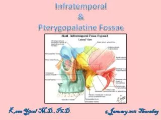
Infratemporal & PterygopalatineFossae • 5.January.2011 Thursday Kaan Yücel M.D., Ph.D.
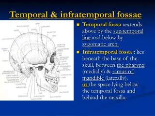
Structures inside thetemporal fossa: • Temporalmuscle • Temporalfascia (overliesthetemporalismuscle) • Superficialtemporalartery (br. of externalcarotid) • Superficialtemporalvein (uniteswiththemaxillaryveinto form theretromandibularvein) • Auriculotemporalnerve (br. of mandibularnervewhich is a br of thetrigeminalnerve)
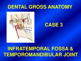
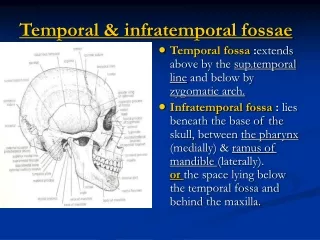
Infratemporal Fossa • Irregularlyshapedspacedeepandinferiortothezygomaticarch, deeptotheramus of themandibleandposteriortothemaxilla. • Communicateswiththetemporal fossa throughtheintervalbetween (deepto) thezygomaticarchand (superficialto) thecranialbones. • Temporal fossa is superiortothezygomaticarch, • Theinfratemporal fossa is inferiortothezygomaticarch.
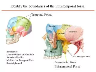
Theboundaries of theinfratemporalfossa • Laterally: ramusof themandible • Medially: lateralpterygoidplate • Anteriorly: posterioraspect of themaxilla • Posteriorly: tympanicplate,mastoidandstyloidprocesses of thetemporal bone • Superiorly: theinferior (infratemporal) surface of thegreaterwing of thesphenoid • Inferiorly: wherethemedialpterygoidmuscleattachestothemandiblenearitsangle
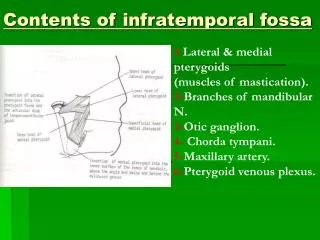
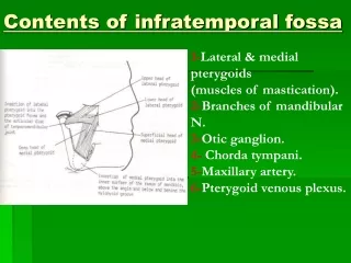
Theinfratemporal fossa containsthe: • Inferiorpart of thetemporalismuscle • Lateralandmedialpterygoidmuscles • Maxillaryartery • Pterygoidvenousplexus • Mandibular, inferioralveolar, lingual, buccal, chordatympaninerves • Oticganglion
Neurovasculature of theinfratemporal fossa • Themaxillaryartery is thelarger of thetwo terminal branches of theexternalcarotidartery. • Itarisesposteriortotheneck of themandibleand is dividedintothreepartsbased on itsrelationtothelateralpterygoidmuscle. • 1st (mandibular) part: Deeptothecondyle of mandible • 2nd (pterygoid) part: Neighbourhood of lateralpterygoidmuscle • 3rd (pterygopalatine) part: Inside theinfratemporal fossa (extendsintothepterygopalatine fossa)
Branches of the 1st part: • Deepauricular (toexternalacousticmeatus) • Anteriortympanicartery (tothetympanicmembrane) • Middlemeningeal (to dura materandcalvaria) • Accessorymeningealaa. (tothecranialcavity) • Inferioralveolarartery (tothemandibulargingivaandteeth)
Branches of the 2nd part: • Deeptemporalaa. (tothetemporalmuscle) • Pterygoidaa. (tothepterygoidmuscles) • Massetericartery (tothemassetermuscle) • Buccalartery (tothebuccinatormuscle) • deepauricular (da) • anteriortympanic (at) • middlemeningeal (mm) • accessorymiddlemeningeal (amm) • inferioralveolar (ia) • buccal (b) • deeptemporal (dt) • posteriorsuperioralveolar (psa) • descendingpalatine (dp) • infraorbital (io) • sphenopalatine (sp)
Pterygoidvenousplexus • Locatedpartlybetweenthetemporalisandthepterygoidmuscles. • Thevenousequivalent of most of themaxillary • Actuallya network of veinsformedbytheveinsfollowingthebranches of maxillaryartery.
Mandibularnerve • Arisesfromthetrigeminalganglion in themiddlecranial fossa. • Immediatelyreceivesthe motor root of thetrigeminalnerve • Leavesthecraniumthroughtheforamen ovale intotheinfratemporal fossa.
Mandibularnerve • Themandibularnervecontains GSA and SVE fibers. • Branches of CN V3 supplythefourmuscles of mastication but not thebuccinator, which is suppliedbythefacialnerve.
Brancheswithintheinfratemporal fossa is dividedintothreegroups: • 1) Branchesarisingfromthetrunk • Spinousnerve • Medialpterygoidnerve • 2) Anterior branches • Buccal nerve • Masseteric nerve • Deep temporal nerves • Lateral pterygoid nerve • 3) Posterior branches • Auriculotemporalnerve • Lingualnerve • Inferioralveolarnerve
Thespinousnervepassesthroughthespinousforamenandentersthecranium. It is a sensorynerveinnervatingthe dura mater. Themedialpterygoidnerveinnervatesthemedialpterygoidmuscle, tensor veli palatinimuscleandthetensortympanimuscle. Buccalnerve, massetericnerve, deeptemporalnerves, lateralpterygoidnerveinnervatethemuscleswiththesame name exceptthebuccalnerve. Buccalnerveis sensoryandinnervatestheinnersurface of thecheek.
Auriculotemporalnerve • Suppliessensoryfiberstotheauricleandtemporalregion. • Alsosendsarticular (sensory) fiberstothe TMJ. • Conveyspostsynapticparasympatheticsecretomotorfibersfromtheoticgangliontotheparotidgland.
Theinferioralveolarnerveentersthemandibularforamenandpassesthroughthemandibularcanal, formingtheinferiordentalplexus, whichsendsbranchestoallmandibularteeth on itsside. Theterminal branch of theinferioralveolarnerve is thementalnervewhichpassesthroughthementalforamen.
Lingualnerve sensorytotheanteriortwothirds of thetongue, thefloor of themouth, andthelingualgingivae.
deeptemporal (dt) • auriculotemporal (at) • inferioralveolar (ia) • nervetothemylohyoid (nmh) • lingual (l) • buccal (b) • branchestolateralpterygoid (not labeled) • Not shown: meningealbranch • nervetomasseter
Chordatympaninerve • A branch of CN VII carryingtastefibersfromtheanteriortwothirds of thetongue. • Joinsthelingualnerve in theinfratemporal fossa. • Alsocarriessecretomotorfibersforthesubmandibular& sublingualsalivaryglands.
Oticganglion(parasympathetic) • Locatedin theinfratemporal fossa, justinferiortotheforamen ovale. Presynapticparasympatheticfibers, derivedmainlyfromtheglossopharyngealnerve(viathelesserpetrosalnerve), synapse in theoticganglion. • Postsynapticparasympatheticfibers, secretorytotheparotidgland, passfromtheoticgangliontothisglandthroughtheauriculotemporalnerve.
auriculotemporalnerve glossopharyngealnerve (viathelesserpetrosalnerve)
Pterygopalatine Fossa • A smallspacebehindandbelowtheorbitalcavity. • An inverted 'tear-drop' shapedspacebetweenbones on thelateralside of theskullimmediatelyposteriortothemaxilla.
Althoughsmall in size, thepterygopalatine fossa communicatesviafissuresandforamina in itswallswith: • themiddlecranial fossa • infratemporal fossa • floor of theorbit • lateralwall of thenasalcavity • oropharynx • roof of the oral cavity
Because of itsstrategiclocation, thepterygopalatine fossa is a major site of distributionforthemaxillarynerve [V2] andforthe terminal part of themaxillaryartery. + parasympatheticfibersfromthefacialnerve [VII] Sympatheticfibersfromthe T1 spinalcordleveljoininingthebranches of themaxillarynerve [V2] in thepterygopalatine fossa. Alltheupperteethreceivetheirinnervationandbloodsupplyfromthemaxillarynerve [V2] andthe terminal part of themaxillaryartery, respectively, thatpassthroughthepterygopalatine fossa.
Skeletal framework The walls of the pterygopalatine fossa are formed by parts of the palatine, maxilla, and sphenoid bones: •anterior wall is formed by the posterior surface of the maxilla; •medial wall is formed by the lateral surface of the palatine bone; •posterior wall and roof are formed by parts of the sphenoid bone.
The part of the sphenoid bone that contributes to the formation of the pterygopalatine fossa is the anterosuperior surface of the pterygoid process. Opening onto this surface are two large foramina: •maxillary nerve [V2] through foramen rotundummiddle cranial fossa •greater petrosal nerve from the facial nerve [VII] + sympathetic fibers internal carotid plexus join to form the nerve of the pterygoid canal that passes into the pterygopalatine fossa through the anterior opening of the pterygoid canal.
The pterygoid canal is a bony canal opening onto the posterior surface of the pterygoid process. • The pterygoid canal opens into the middle cranial fossa just anteroinferior to the internal carotid artery as the vessel enters the cranial cavity through the carotid canal.
The contents of the pterygopalatinefossa 1) Third part (pterygopalatine part) of the maxillary artery 2) Maxillary nerve 3) Nerve of the pterygoidcanal (Vidian’snerve) Pterygopalatineganglion
Lacrimal Gland • Consistsof a large orbital part and a small palpebral part. • Situatedabove the eyeball in the anterior and upper part of the orbit posterior to the orbital septum. • Opens into the lateral part of the superior fornix of the conjunctiva.
Innervation of the lacrimal gland The parasympathetic secretomotor nerve supply is derived from the lacrimal nucleus of the facial nerve. Thepreganglionicfibersreachthepterygopalatineganglionviathenervusintermediusanditsgreatpetrosalbranch.
The postganglionic parasympathetic and sympathetic fibers leave the zygomaticotemoral branch of the zygomatic nerve and form a special autonomic nerve, which joins the lacrimal nerve. • The lacrimal nerve is a major general sensory branch of the ophthalmic nerve [V1]. • The postganglionic parasympathetic and sympathetic fibers pass with the lacrimal nerve to the lacrimal gland.
Maxillary Nerve (V2) • Purelysensory • Originatesfrom the trigeminal ganglion in the cranial cavity • Exitsthe middle cranial fossa • Enters the pterygopalatine fossa (foramen rotundum) • Exits as the infra-orbital nerve (inferior orbital fissure) • Gives sensory fibers to the skin of the face and the side of the nose.
Branches • Within the fossa, the maxillary nerve is attached to the pterygopalatineganglionby two ganglionic branches. • Sensoryfibers from the nose, the palate, and the pharynx. • Postganglionicparasympathetic fibers to the lacrimal gland.
More anteriorly posterior superior alveolar nerves are given off. Pass through the pterygopalatine maxillary fissure into the infratemporal fossa.
Here they divide into numerous small branches • Enterthe maxilla through the posterior alveolar foramina • Supplythe upper molar teeth, the mucous membrane on the buccal surface of the associated alveolar process and the lining of the maxillary sinus. Anesthesia of the upper molar teeth and associated buccal mucosa can be achieved by a posterior superior alveolar block.
As the maxillary nerve is about to enter the inferior orbital fissure it gives rise to the zygomatic nerve. divides into: Zygomaticotemporalbranch passing into temporal fossa to supply skin of the temple Zygomaticofacialnerve supplies skin over the prominence of cheek.
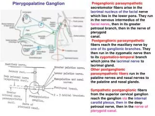
PterygopalatineGanglion • (Ganglionpterygopalatinum, Meckel'sganglion, Nasalganglion, Sphenopalatineganglion) • Largestof the 4parasympathetic ganglia in the head • Formedby the cell bodies of the postganglionic neurons associated with preganglionic parasympathetic fibers of the facial nerve [VII] carried by the greater petrosal nerve and the nerve of the pterygoid canal.
Pterygopalatine Ganglion • The postganglionic fibers, together with sympathetic fibers, join fibers from the ganglionic branches of the maxillary nerve [V2]. • Postsynaptic fibers arising from the pterygopalatine ganglion supply the lacrimal gland as well as nasal glands and minor salivary glands within the oral cavity. The postsynaptic fibers innervating the lacrimal gland pass to the lacrimal nerve to reach the lacrimal gland.
Branches •Orbital branches, enter the orbit (inferior orbital fissure) Supplyof the orbital wall and of the sphenoidal and ethmoidal sinuses. •Greater and lesser palatine nerves, supply the palate, the tonsil, and the nasal cavity. The greater palatine nerve originates from the geniculate ganglion of the facial nerve [VII] in the temporal bone. In the palatine canal, gives origin to posterior inferior nasal nerves, which contribute to the innervation of the lateral nasal wall. Greater petrosal nerve enters the pterygoid canal and becomes the nerve of the pterygoid canal •Pharyngeal branch, which supplies the roof of the nasopharynx
infraorbitalnerve posteriorsuperioralveolarnerve pterygopalatineganglion (parasympathetic) greaterpalatinenerve lesserpalatinenervecut nasopalatinenerve nerveof thepharyngealcanal
Maxillary Artery • Amajor branch of the external carotid artery in the neck. • Passes through the infratemporalfossa • Entersthe pterygopalatine fossa through the pterygomaxillary fissure.