Download
1 / 14
210 likes | 548 Vues
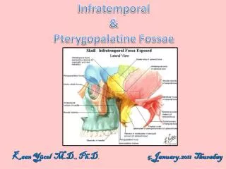
PTERYGOPALATINE FOSSA. By.pullanna . Dept.of anatomy,ymc. The Pterygopalatine fossa. Inverted 'tear-drop' shaped space Between bones on the lateral side of the skull Immediately posterior to the maxilla Small in size. Sphenoid bone.

E N D
PTERYGOPALATINE FOSSA By.pullanna . Dept.of anatomy,ymc.
The Pterygopalatine fossa • Inverted 'tear-drop' shaped space • Between bones on the lateral side of the skull • Immediately posterior to the maxilla • Small in size
Sphenoid bone • The part of bone that contributes to the formation of the fossa is the anterosuperior surface of the pterygoid process • Opening onto this surface are two large foramina: • 1.The Foramen rotundum • 2.Pterygoid canal
Foramen rotundum • Lateral and superior foramen • Communicates posteriorly with the middle cranial fossa • The maxillary nerve [V2] passes through it
Contents • 1. The maxillary nerve [V2] • 2. Terminal part of the maxillary artery • 3. Nerve of the pterygoid canal • 4. The pterygopalatine ganglion • 5. Veins and lymphatics also pass through the pterygopalatine fossa.
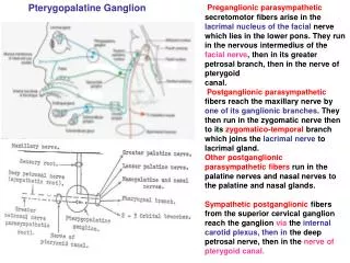
Pterygopalatine ganglion • Largest of the four parasympathetic ganglia in the head • Formed by the cell bodies neurons associated with: • 1. Preganglionic parasympathetic fibers of the facial nerve carried by the greater petrosal nerve and the nerve of the pterygoid canal. • 2.Sensory and ganglionic branches of the maxillary nerve • 3.Postganglionic sympathetic fibers (deep petrosal)
Motor or parasympathetic root: • Secretomotor fibers to the lacrimal gland. • Glands of the nose. • Palate. • Nasopharynx. • Para nasal sinuses. Sympathetic root:provide vasomotor supply • The mucous membrane of the nose. • Palate. • Pharynx.
Branches • Orbital inferior orbital fissur • Palatine • Nasal sphenopalatine foramen • Pharyngeal palato vaginal canal