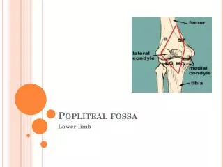
POPLITEAL FOSSA
300 likes | 554 Vues
Learn about the boundaries, structures, and contents of the popliteal fossa, including the popliteus muscle and popliteal vessels. Find detailed information and diagrams on this crucial anatomical region.

POPLITEAL FOSSA
E N D
Presentation Transcript
POPLITEAL FOSSA https://rad.washington.edu/muscle-atlas/popliteus/
ROOF • Skin, • Superficial fascia, • Popliteal fascia , pierced by : Short Saphenous vein, • Post cutaneous nerve of thigh • Superficial contains:- • Short saphenous vein • Post cut nerve of thigh • Post div of medial cut nv • of thigh • Sural communicating nv
Popliteus Muscle • Origin inferior, popliteal surface of tibia, above the soleal line, fascia of semimembranosus • Deep to arcuatepopliteal ligament • Enters capsule • Crosses lateral surface of lateral meniscus • Attached by popliteal-meniscal fibres which bound hiatus • Enters hiatus • Crosses femoral condyle • Deep to lateral collateral ligament • Inserts into anterior part of groove • Superior popliteal recess communicates joint
Origin of the pupl. muscle • The popliteus muscle has three origins, the strongest of which is from the lateral femoral condyle. Other important origins are from the fibula (popliteofibular ligament) and from the posterior horn of the lateral meniscus. The femoral and fibular origins form the arms of an oblique Y-shaped ligament, the arcuate. The arms are joined together by the capsule and meniscal origin.
POPLITEALFOSSA • Termination of Short Saphenous Vein • Contents : • Popliteal vessels • Tibial nerve • Comn peroneal nerve • Popliteal Lymph nodes • Fat • Genicular Branch of Post. Divn. of Obturator nerve • Post cutanous nerve of the thigh ( before it becomes cutaneous) • Sural communicating nerve
POPLITEAL FOSSA :Contents • Popliteal Artery: • From Adductor Hiatus to Lower Border ofPopliteus • Passes Dwn & lat along thefloor • Div into Ant & post tibialarteries
POPLITEAL VESSELS & TIBIALNERVE Anterior Medial Lateral UPPER MIDDLE LOWER Posterior
Popliteal Artery -Contd • Branches : • Cutaneous : Skin of back of leg • Muscular : • Articular : Five Genicular arteries • Sup & Inf Med , Sup & Inf Lat, • Middle • Anastomosis around Knee • Desc Br of Lat Cx Femoral, (SL) • Desc genicular br of femoral (SM) • Sup medial genicular & Saphenous br of desc genicular art (IM) • Rec brs of Ant tibial , Cx Fibular br of Post tibial (IL)
POPLITEAL FOSSA :Contents • Popliteal Nodes: • 6 in no • Afferents: Superficial lymphatics acc SS Vein, (Postero lateral aspect of leg & foot) • From Knee joint • Deep lymphatics acc Tibial vessels. • Efferents: Acc Femoral vessels to Deep Inguinal nodes
Proximal Tibiofibular Joint Articulation is between the lateral condyle of the tibia and the head of the fibula.The articular surfaces are flattened and covered by hyaline cartilage. This is a synovial, plane, gliding joint. Ligaments Anterior and posterior ligaments strengthen the capsule. The interosseous membrane Capsule and Synovial Membrane attached to the line of the articular surface. The common peroneal nerve supplies the joint. Movements A small amount of gliding movement takes place during movements at the ankle joint.
Distal Tibiofibular Joint Articulation Articulation is between the fibular notch at the lower end of the tibia and the lower end of the fibula. Type The distal tibiofibular joint is a fibrous joint. Capsule There is no capsule