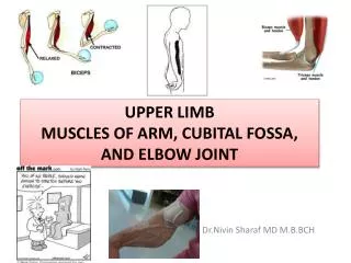
Cubital fossa
Cubital fossa. M y B um T urns R ed (from med to lat) M edian nerve B rachial artery T endon of bicep R adial nerve. Carpal Tunnel- Learn this backwards. . Roof flexor retinaculum fibrous band Floor & walls carpus Contents flexor tendons & median nerve

Cubital fossa
E N D
Presentation Transcript
Cubitalfossa • My Bum Turns Red (from med to lat) • Median nerve • Brachial artery • Tendon of bicep • Radial nerve
Carpal Tunnel- Learn this backwards. • Roof flexor retinaculum fibrous band • Floor & walls carpus • Contents flexor tendons & median nerve median nerve
Boundaries of the Axilla • APEX: • Clavicle • Coracoid process • 1st rib • BORDERS: • Anterior: pectoralis major & minor • Posterior: lats dorsi, teres major, subscapularis • Medial: serratus anterior, ribs, intercostals • Lateral: tendon of long head of biceps brachii • BASE: • Axillary fascia • Runs between latissimus dorsi and pectoralis major • ie. Hairy armpit
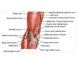
The cubital fossa is the triangular area on the anterior view of the elbow • Boundaries • superior (proximal) boundary - an imaginary horizontal line connecting the medial epicondyle of the humerus to the lateral epicondyle of the humerus • medial (ulnar) boundary - lateral border of pronator teresmuscle • lateral (radial) boundary - medial border of brachioradialis muscle • apex- formed by the meeting point of the lateral and medial boundaries • Contents • The cubital fossa contains four main vertical structures (from lateral to medial): • The radial nerve • The biceps brachii tendon • The brachial artery. (before splitting into radial and deeper ulna artery) • The median nerve • The ulnar nerve is also in the area, but is not in the cubital fossa; it occupies a groove on the posterior aspect of the medial epicondyle of the humerus.